E-Case report
Burst Fracture L1
Posterior fixation T11 – L3 Christoph Grimm, MD


Pre OP

Clinical Case – Burst Fracture L1
Posterior fixation T11 – L3
Neo ADVISE™
Trauma module, Marker Detection
Dr. Christoph Grimm
Neurosurgeon
Bezirkskliniken Schwaben
Standort Günzburg
Günzburg, Germany


Patient Information:
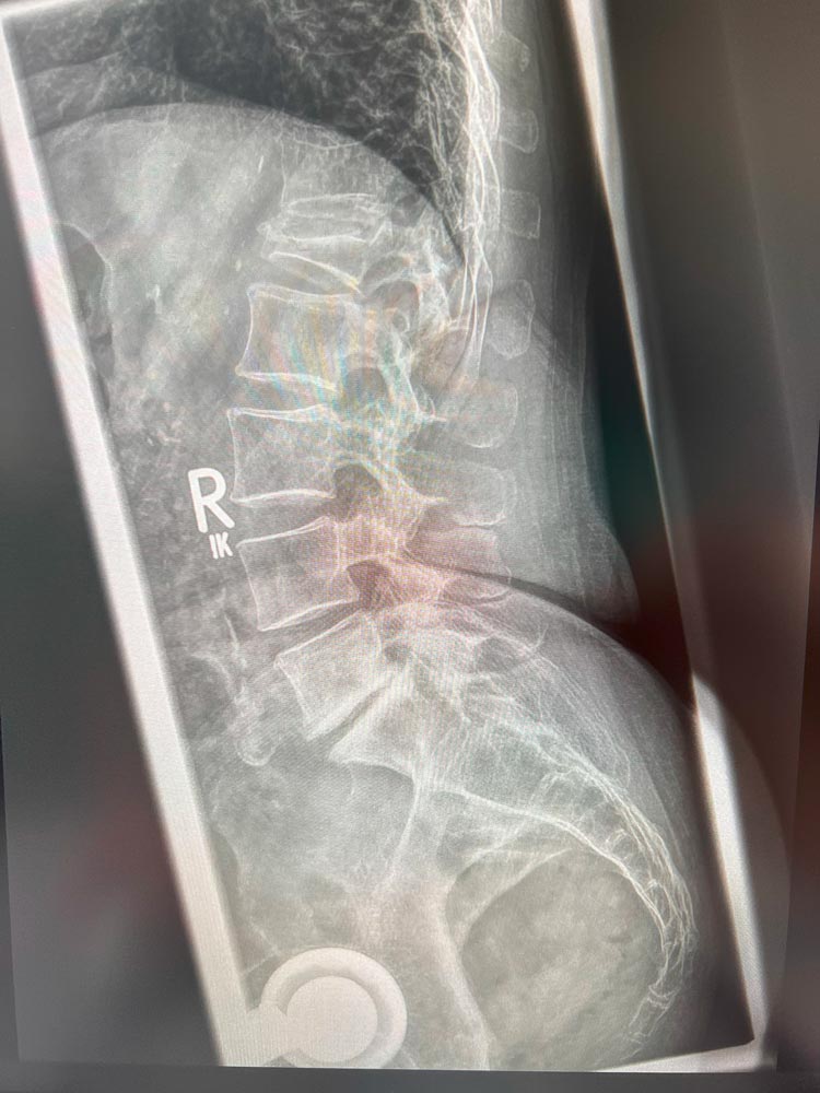
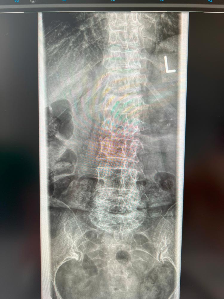
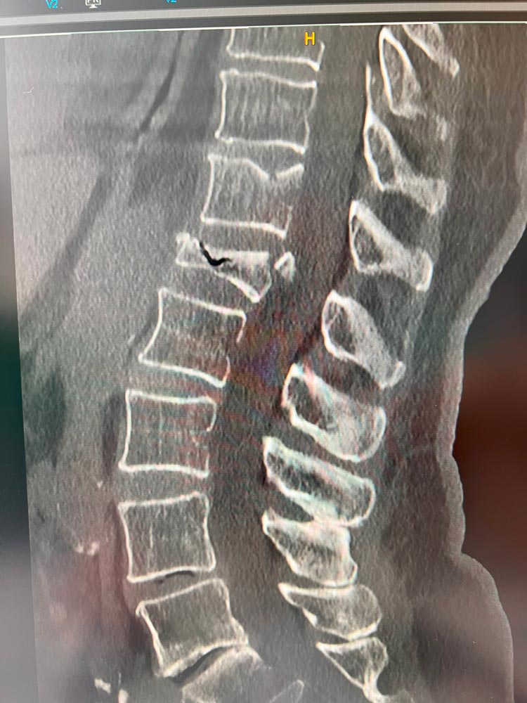
79-year-old female patient suffered a burst fracture in L1 level
- A4 type fracture involving the posterior wall, fragmented
- Spinal stenosis at the fractured level
- Compromised bone quality
Planned Surgery:
- Percutaneous MIS surgery, lateral approach
- Midline approch for navigation star (Brainlab Navigation)
- Posterior fixation T11 – L3
- Intermediate screws in the fracturd level L1
- Cement augmented screws T11 and L3
- Neo ADVISE™ Marker Detection Method to be used


Pre OP radiographs & CT, sagittal and frontal views

Intra OP
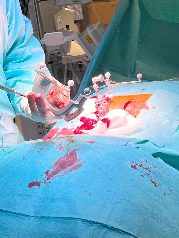
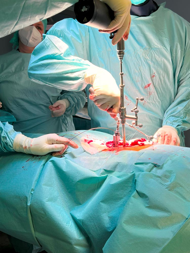
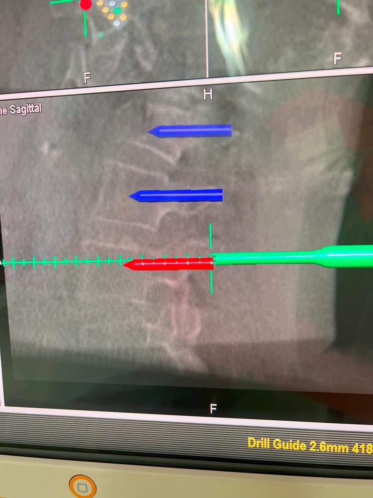
Lateral access incisions, and midline access for the navigation star, Brainlab drillguide
Navigating and placing the first K-wire


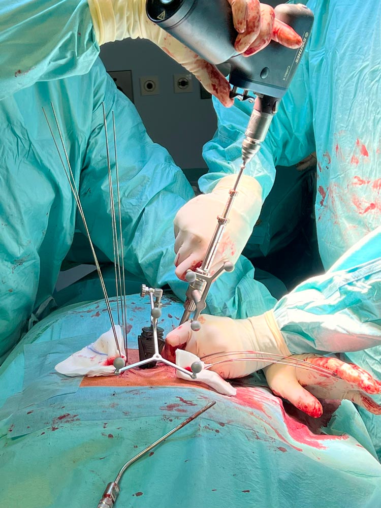
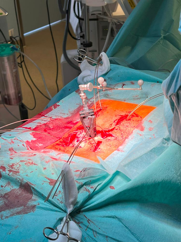
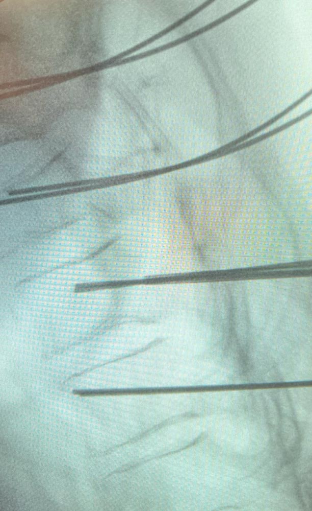
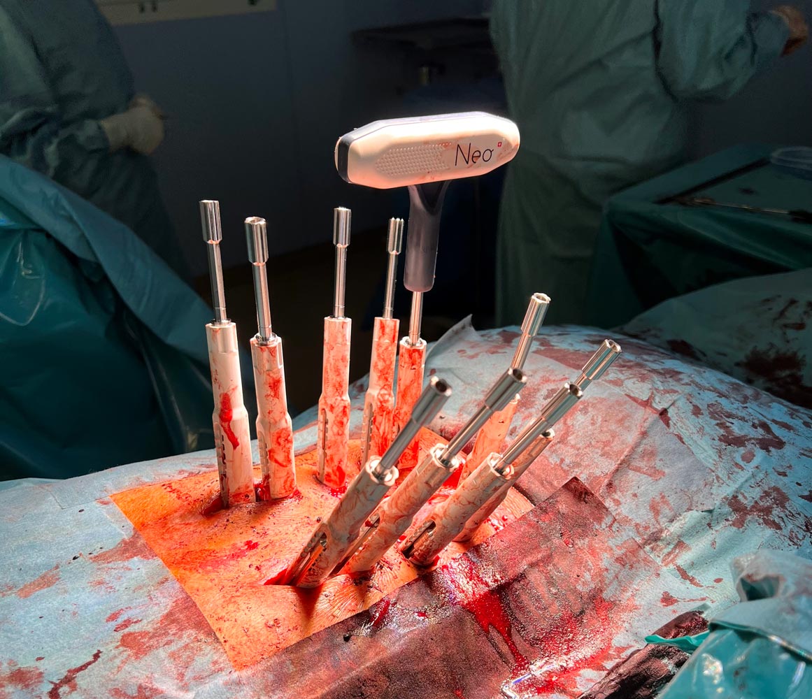
All 10 K-wires have been placed


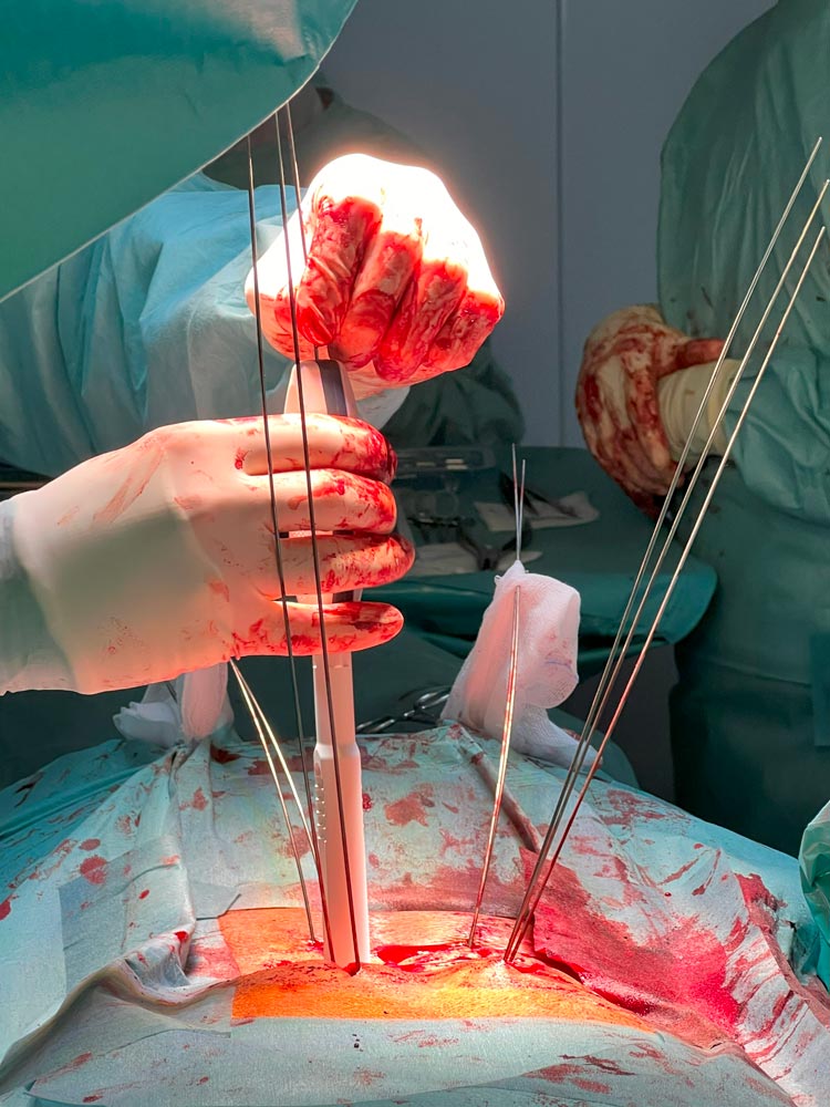
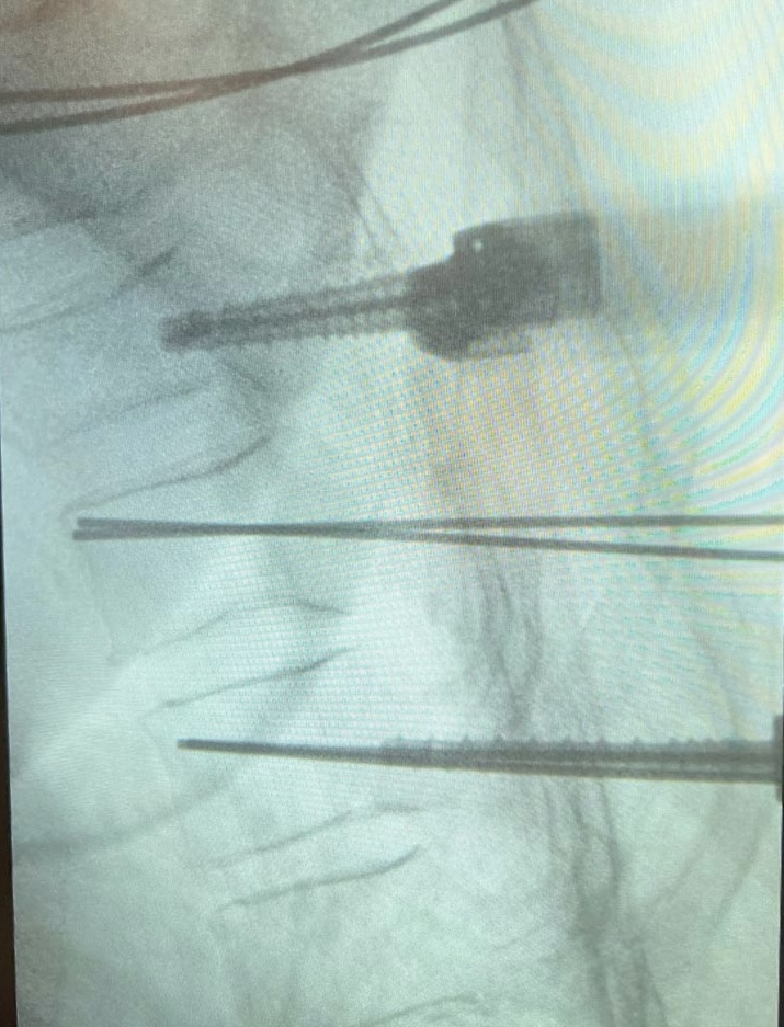
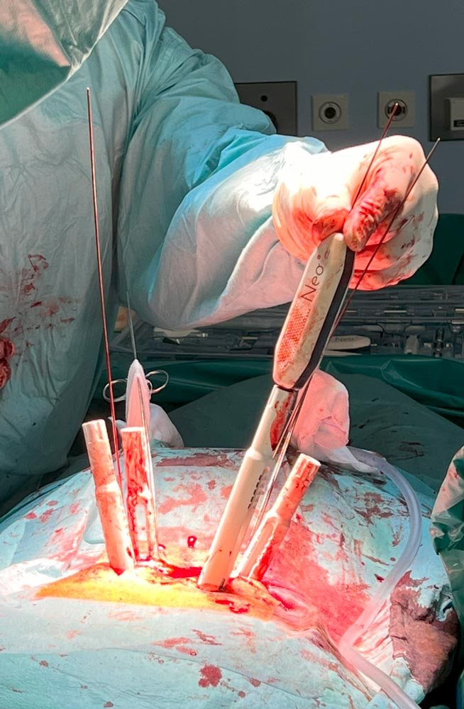
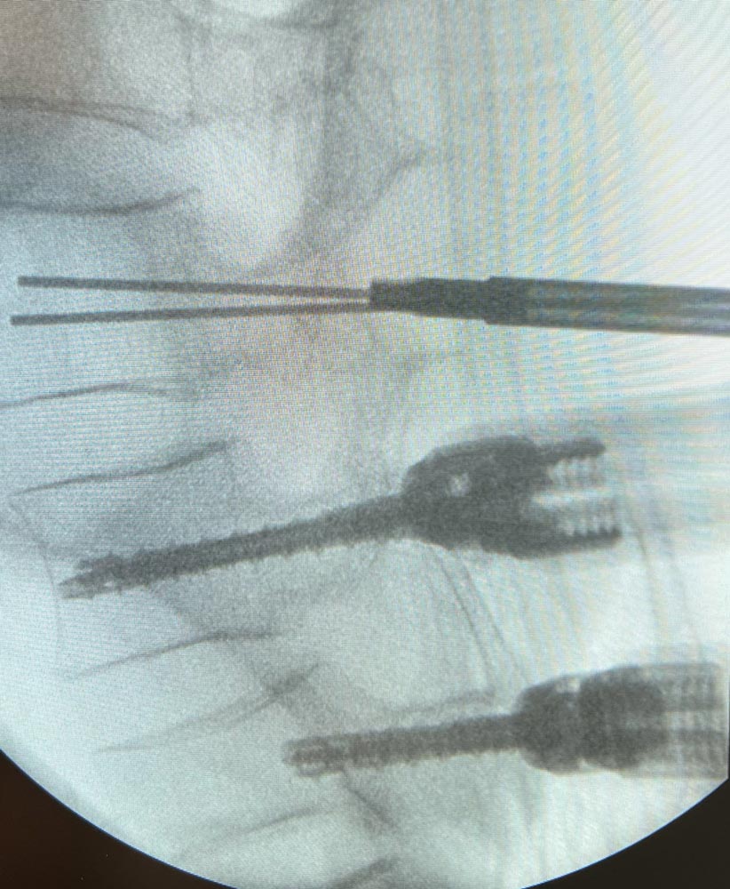
Posterior fixation with Neo Pedicle Screw System™. Starting the pedicle screws insertion


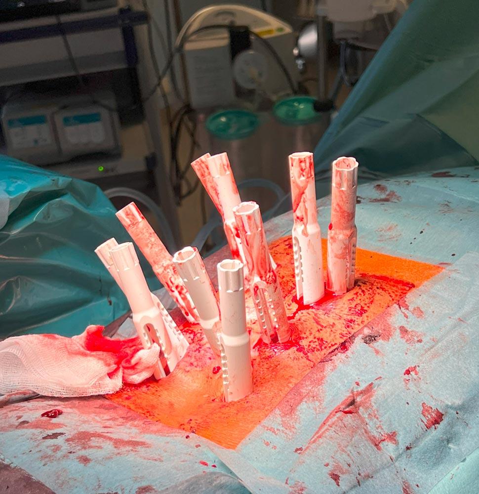
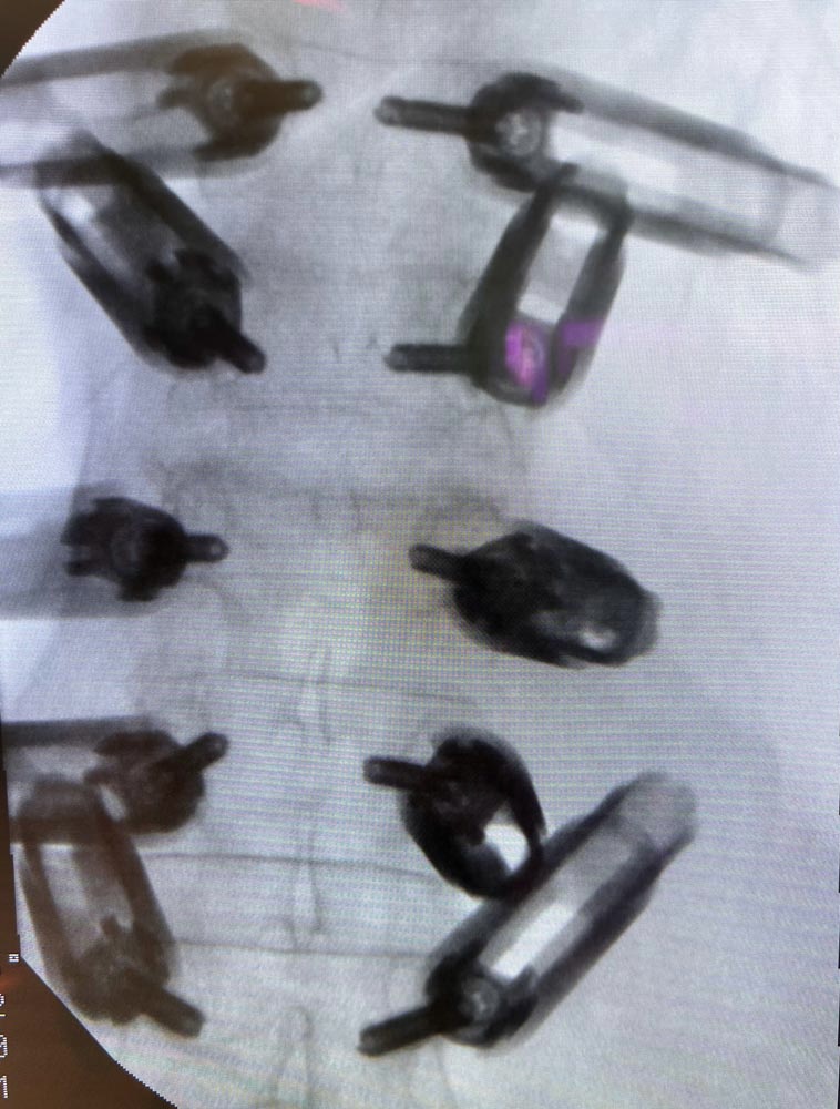
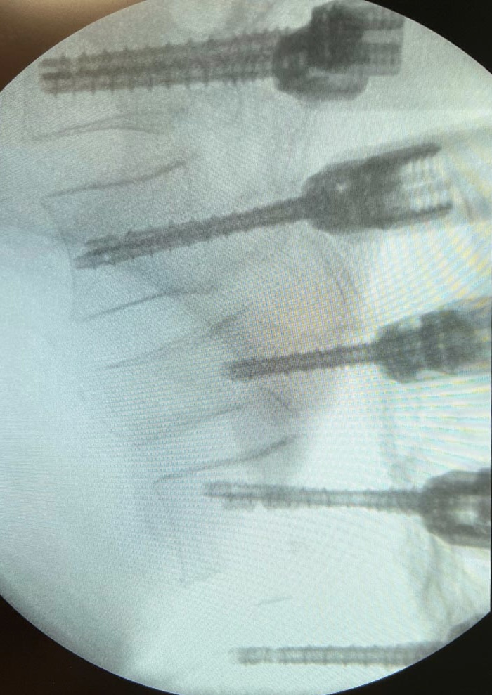
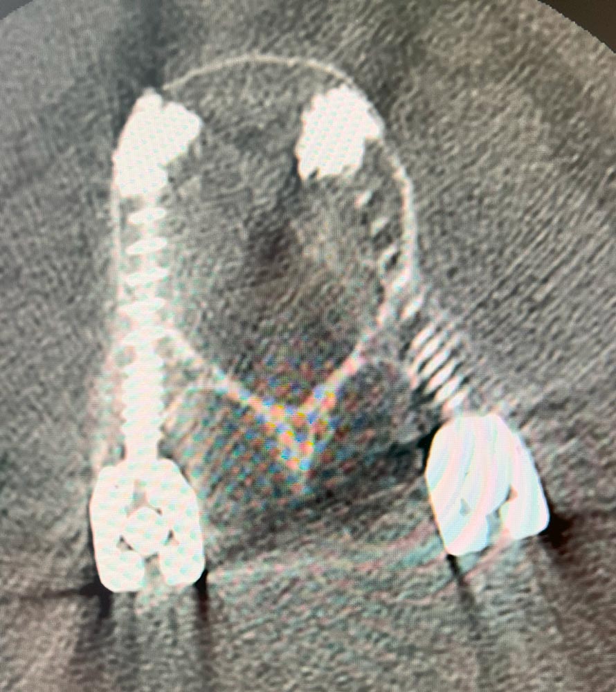
Totally 10 pedicle screws were placed over the levels T11 – L3
Intermediate shorter screws in the fractured L1
- T11 – 2 x Ø5.0x45 mm polyaxial
- T12 – 2 x Ø5.0x45 mm polyaxial
- L1 – 2 x Ø5.0x35 mm polyaxial
- L2 – 2 x Ø5.0x50 mm polyaxial
- L3 – 2 x Ø6.0x50 mm polyaxial

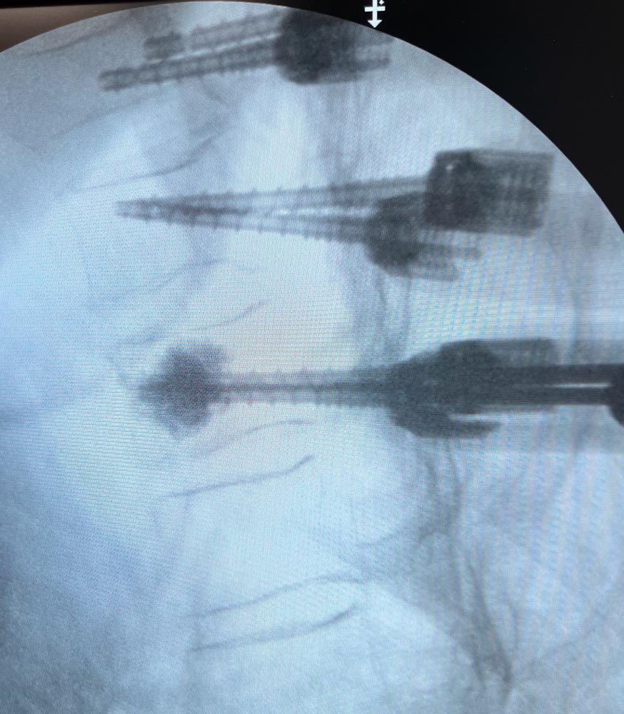
Cement augmentation of pedicle screws in levels T11 and L3, 1ml cement/screw.
All Neo system pedicle screws are cannulated and fenestrated to faciliate this surgical step.
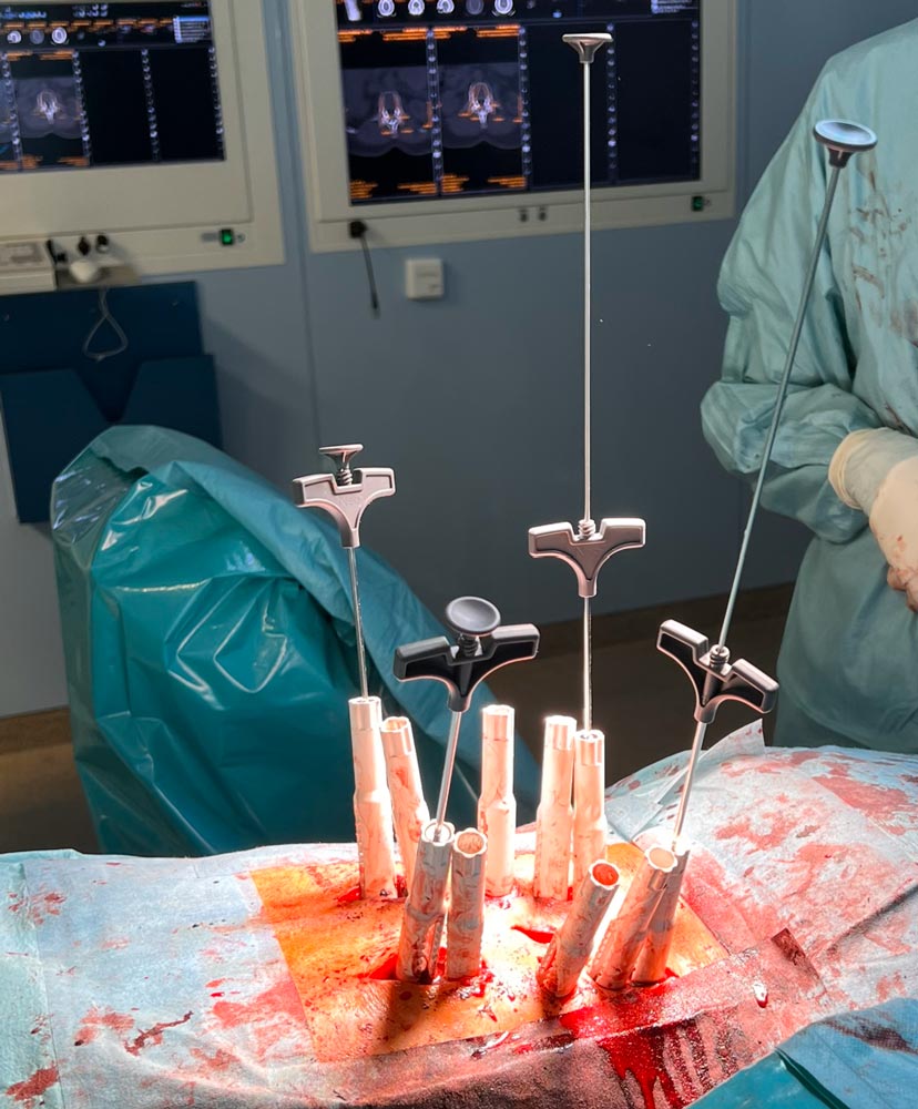
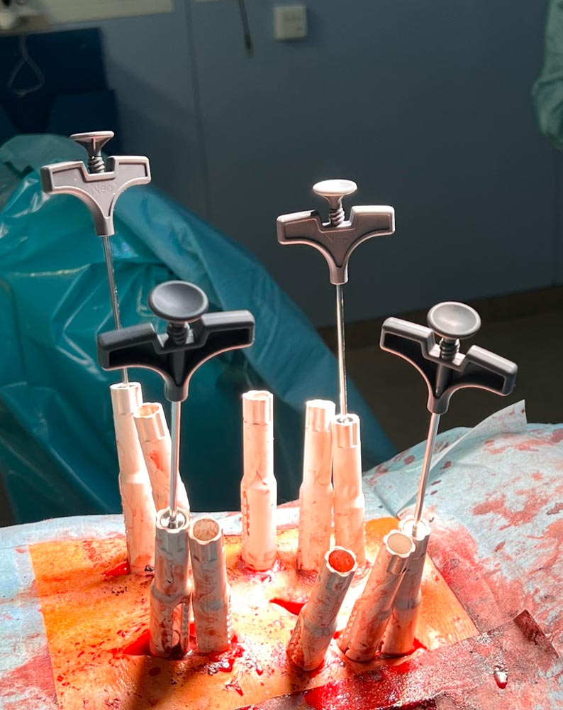
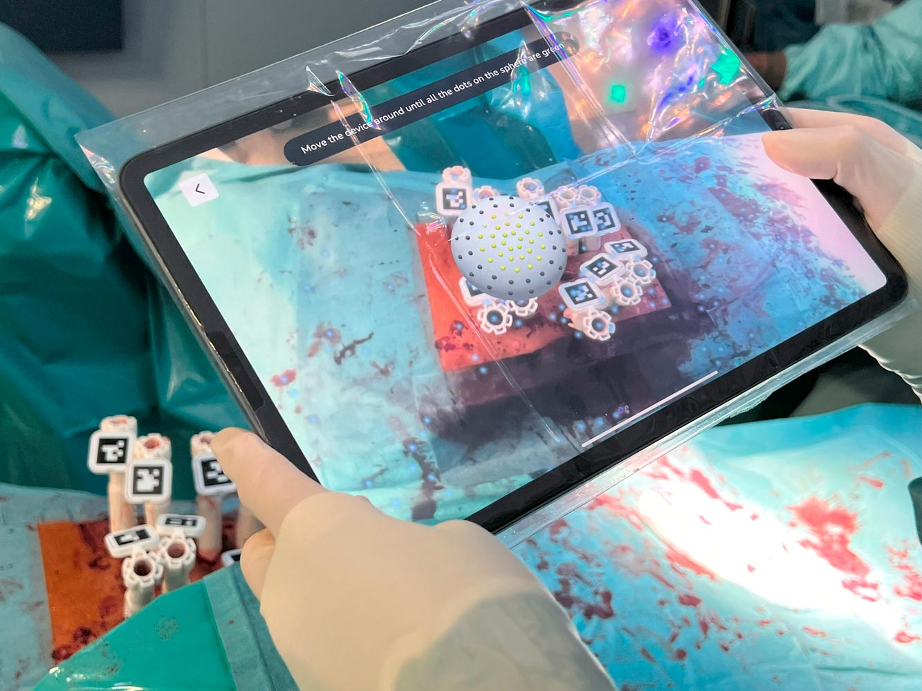
Usage of Neo ADVISE™ Marker Detection Method
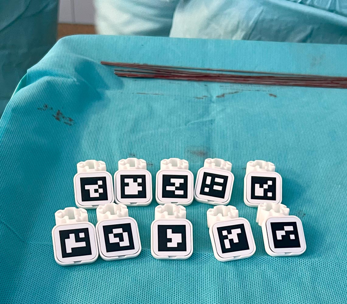
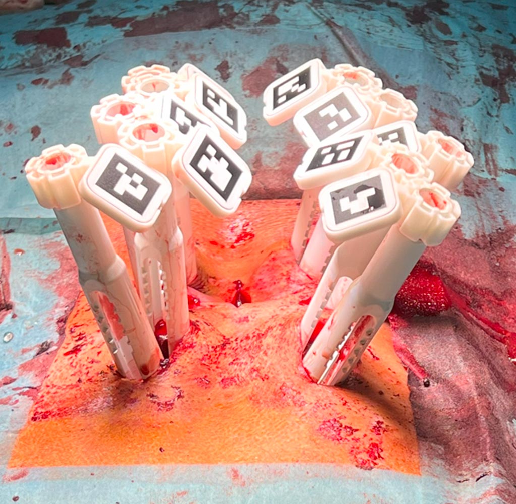
The QR code Markers are attached to the guide towers
The Scene Analysis is performed

Finding and mapping the Markers
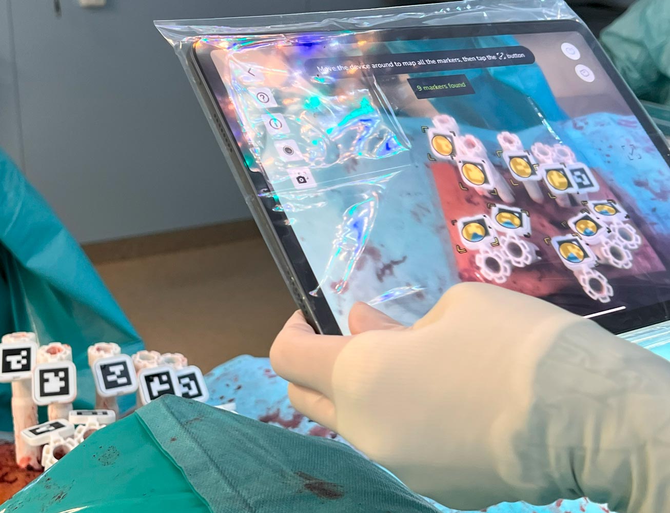
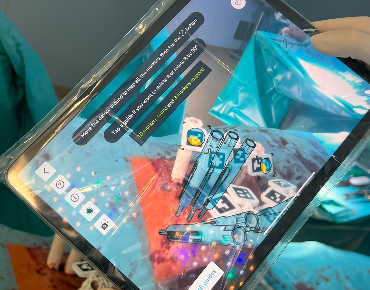


Results from using the Neo ADVISE™ in both the left and the right side
Visualizations of the rods in both the sagittal and the coronal planes in the iPad screens
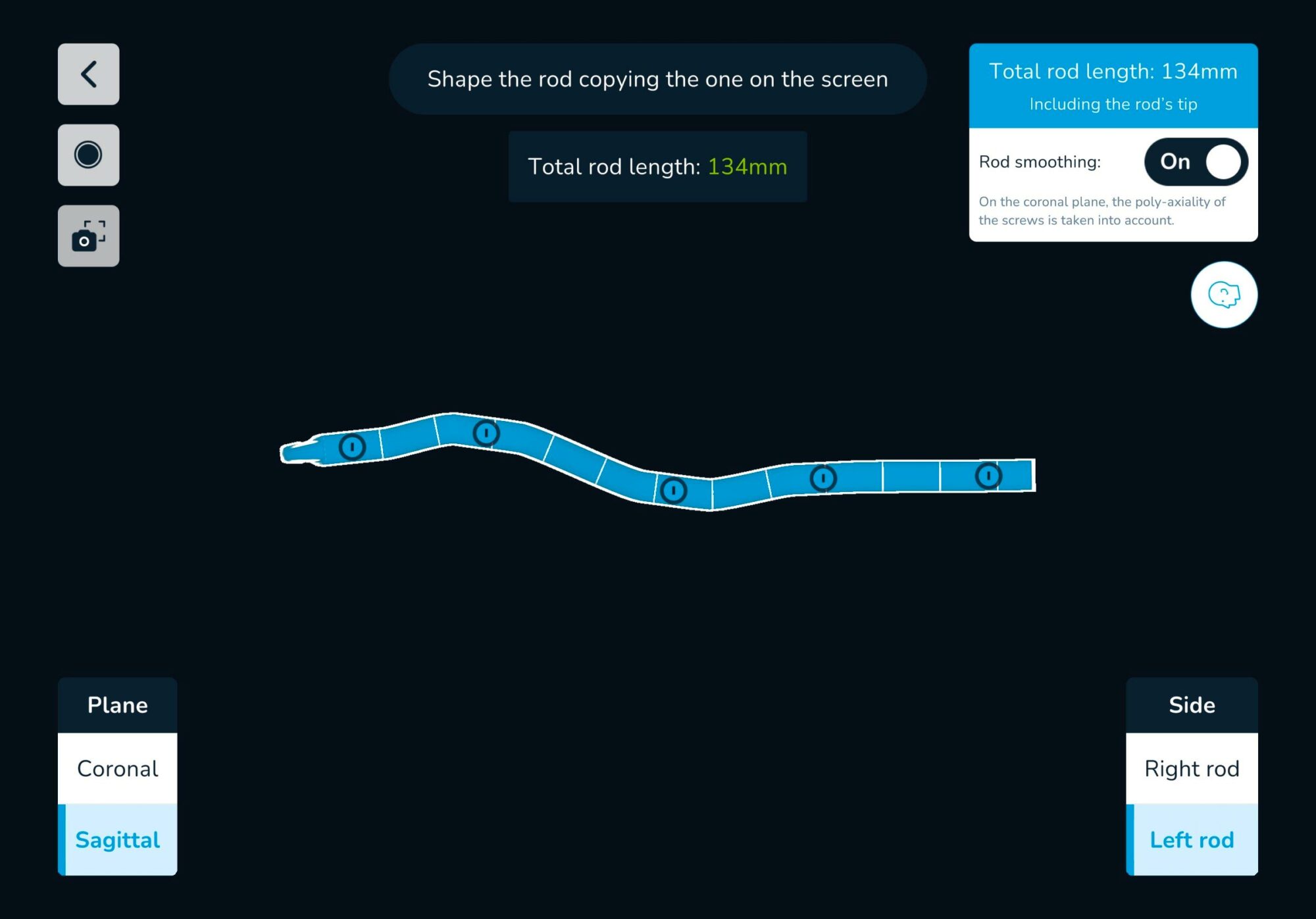
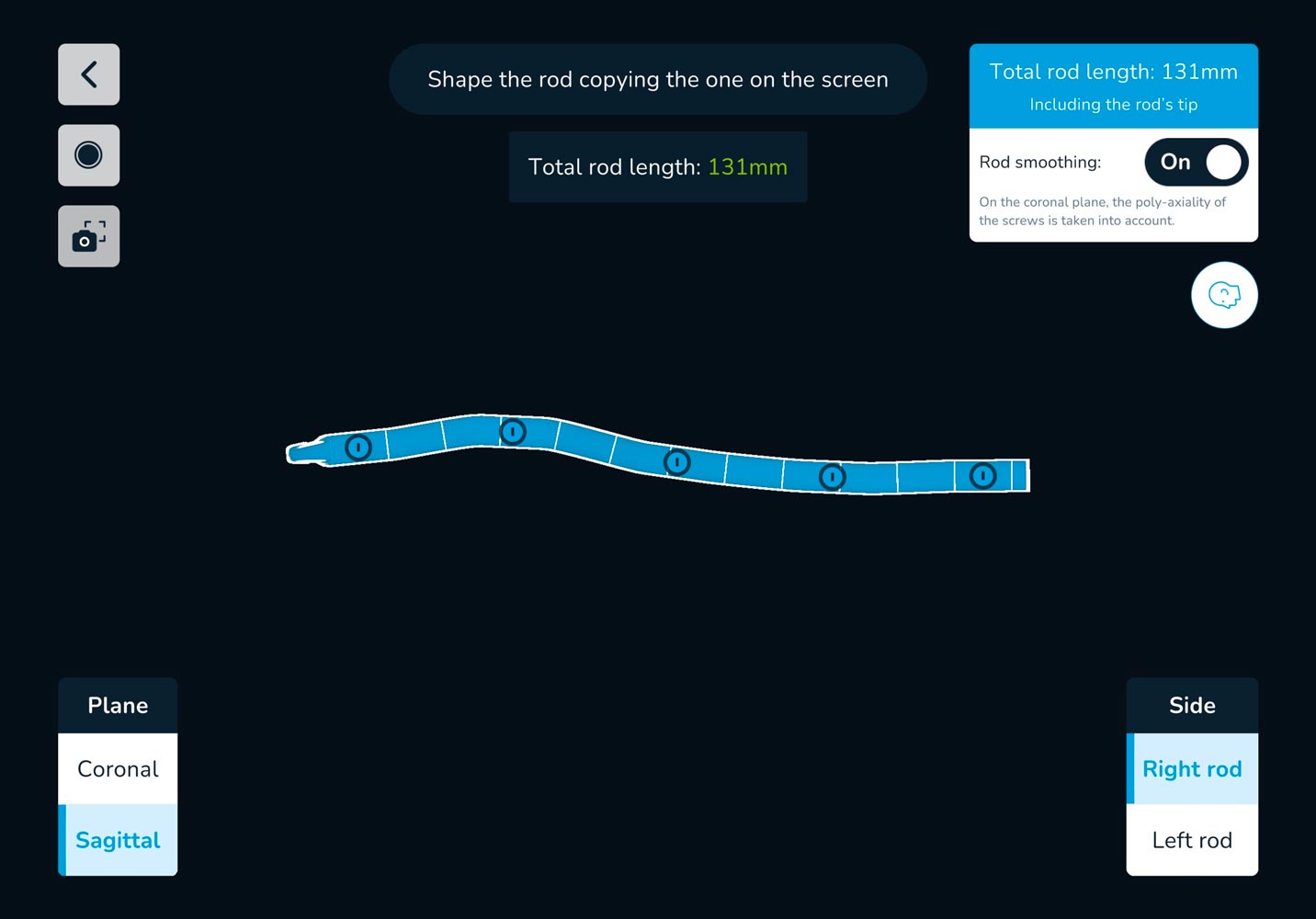
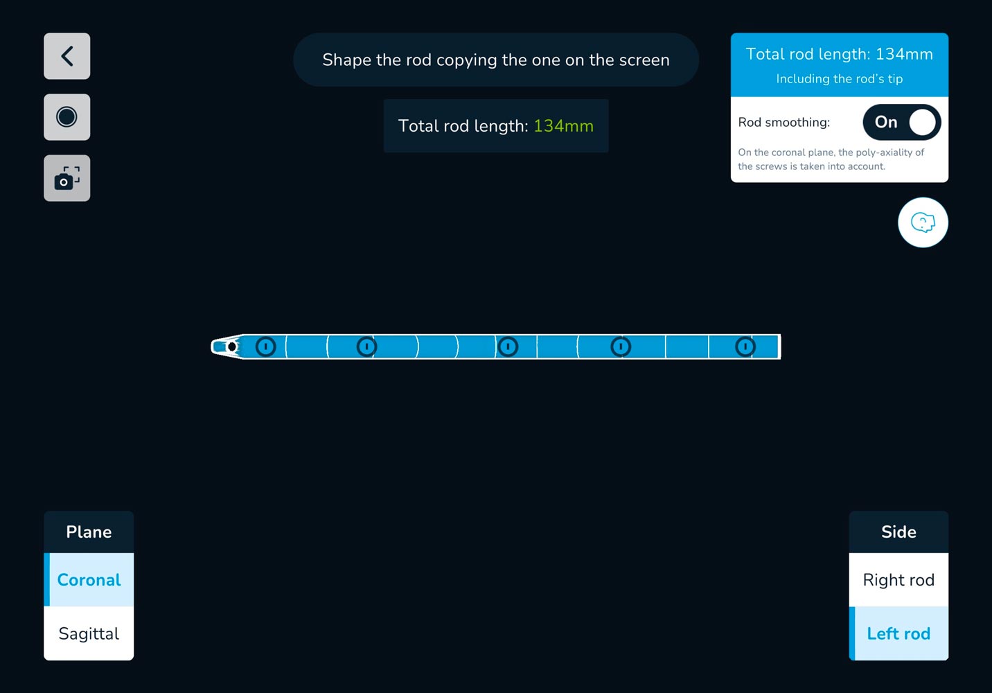
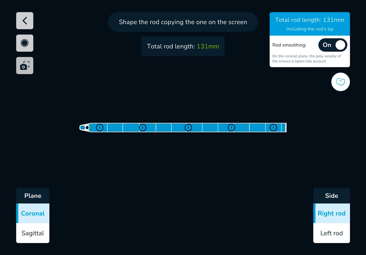
Rod selection: 2 x Titanium rods 160mm straight, shortened to 135mm


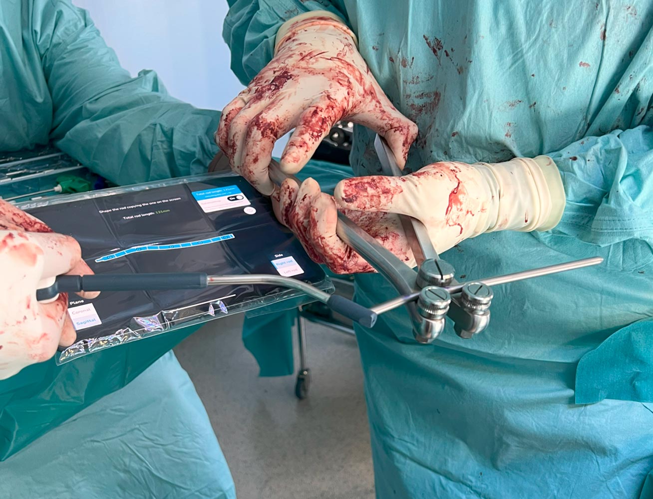
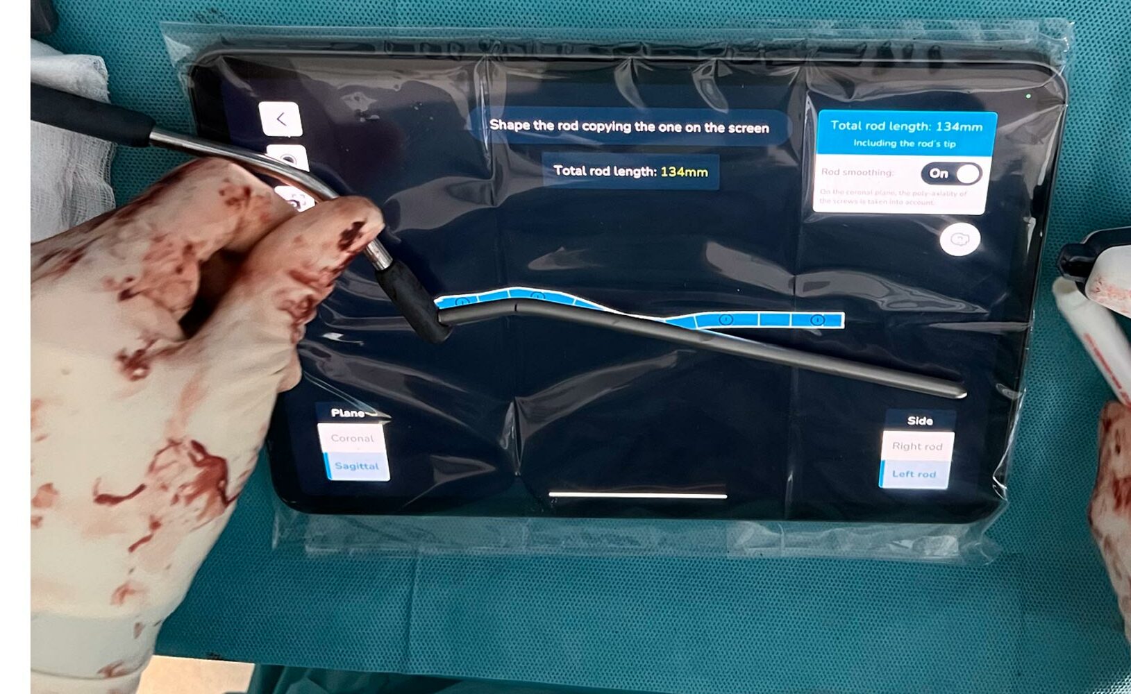
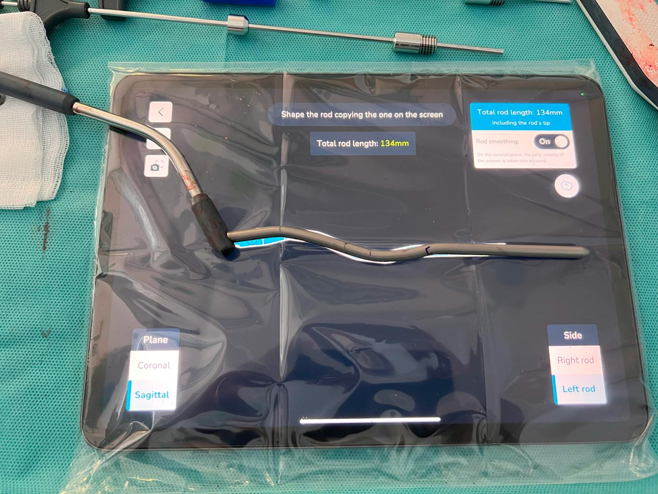
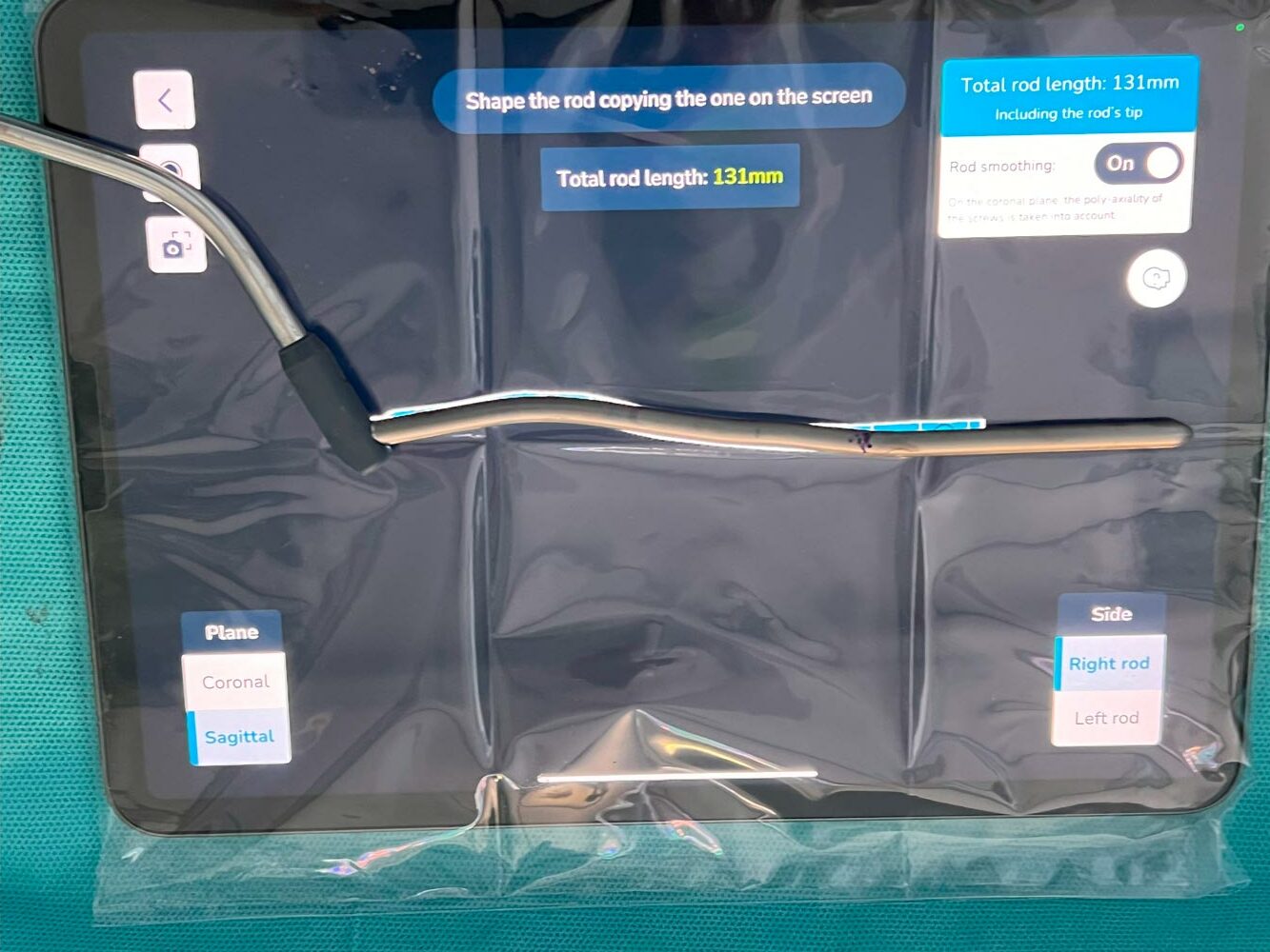
Rod bending using the rod visualization in the iPad screen as template for the sagittal shape of both rods.


Rod placement & Final set screw tightening
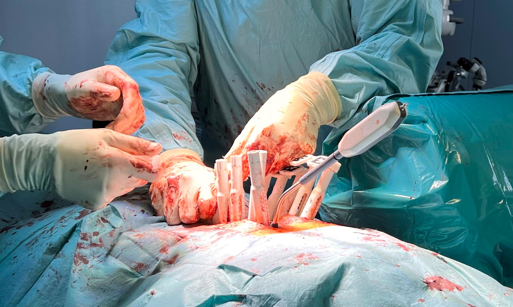
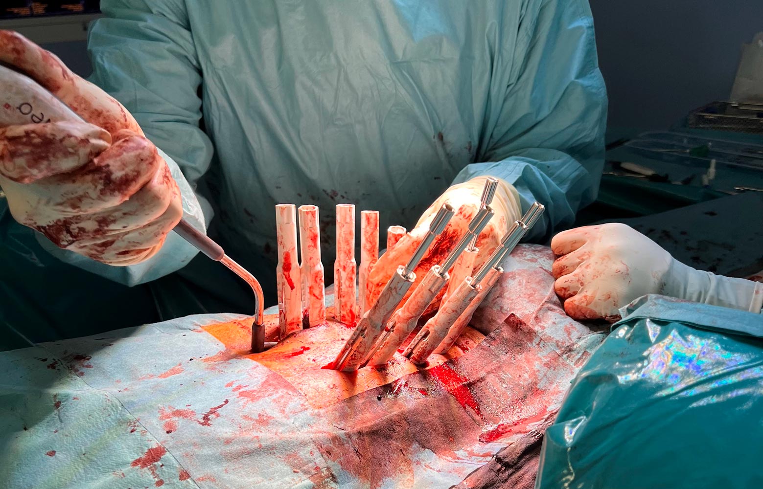

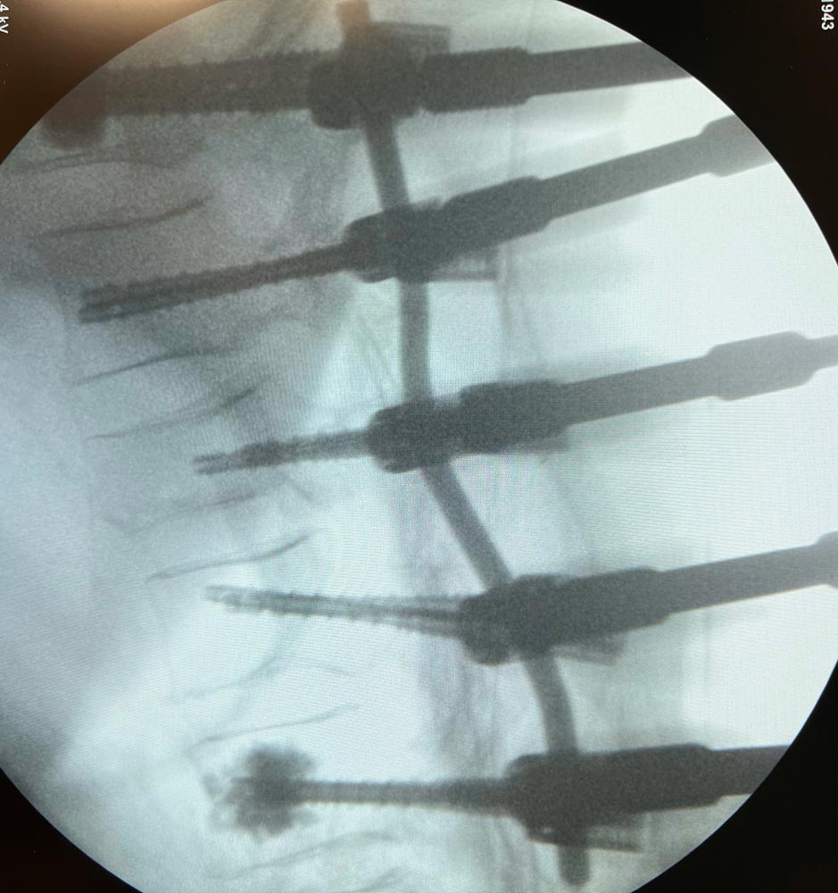
Rod selection: 2 x Titanium rods 160mm straight, shortened to 135mm


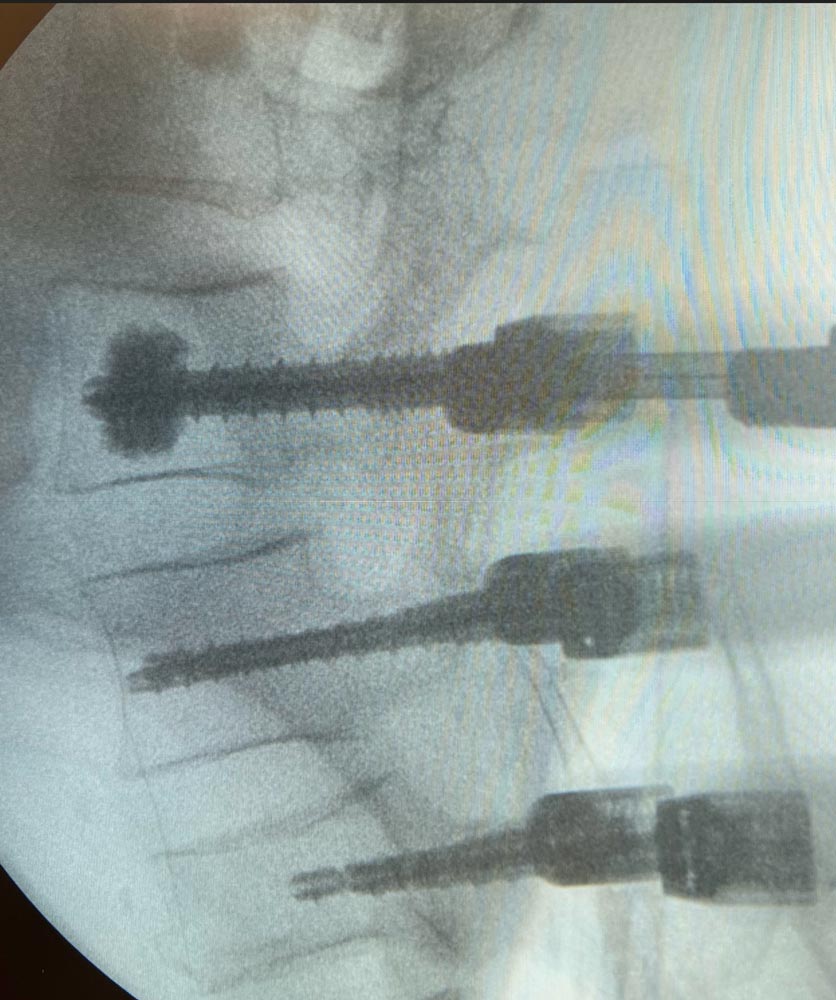
Radiographs after final fixation of the posterior construction and removal of the instrumentation
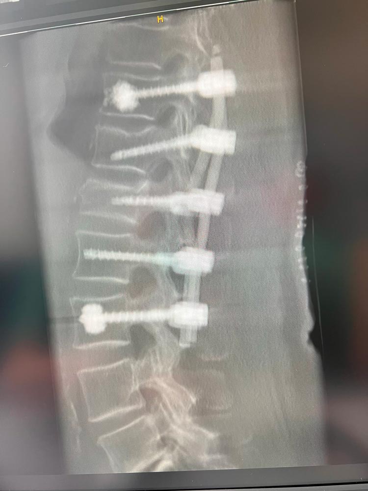
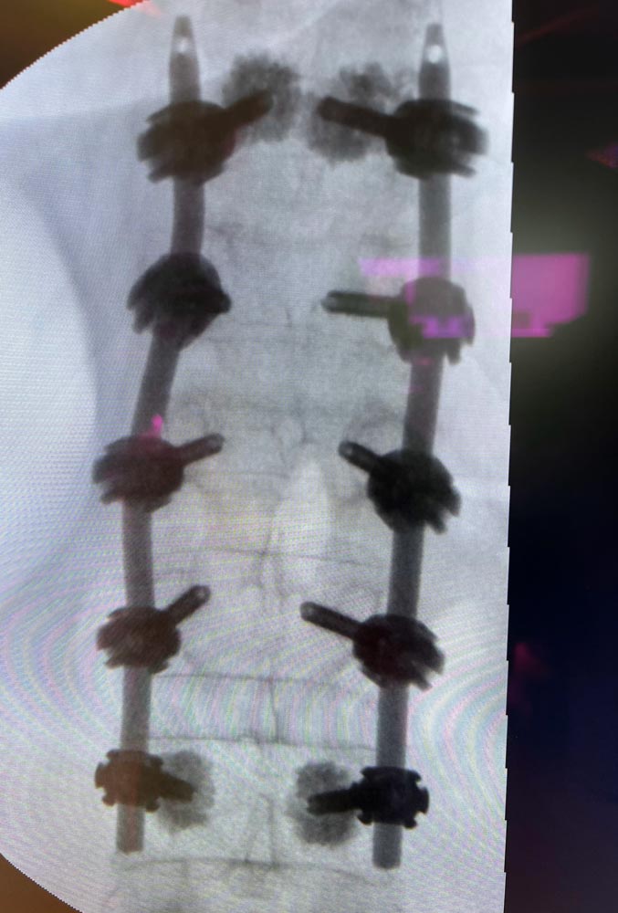
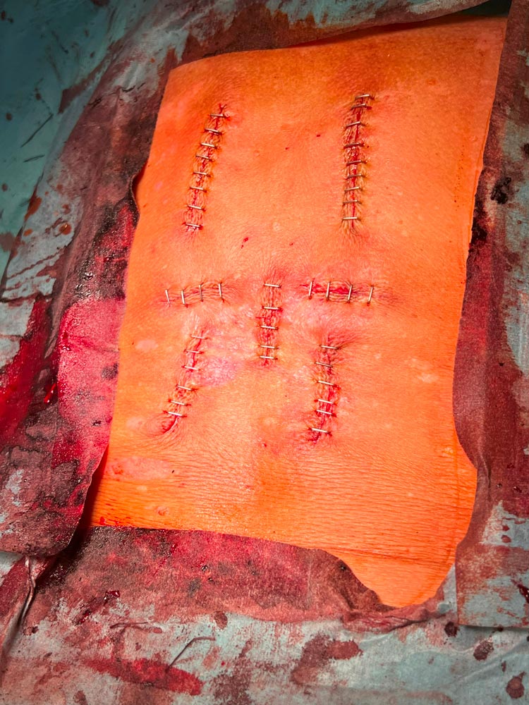
Closure of incision

Post OP
Radiographs after the final fixation of the posterior construction, and removal of the instrumentation

Post OP CTs
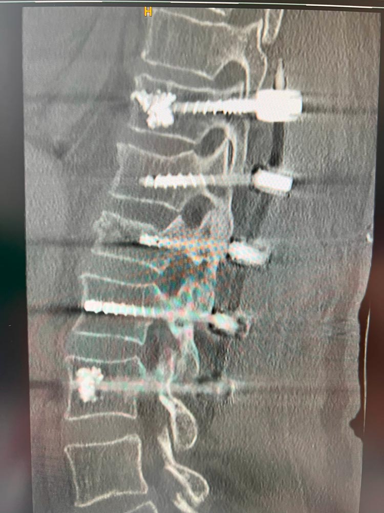
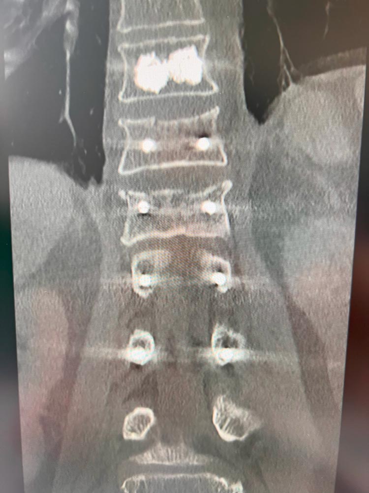
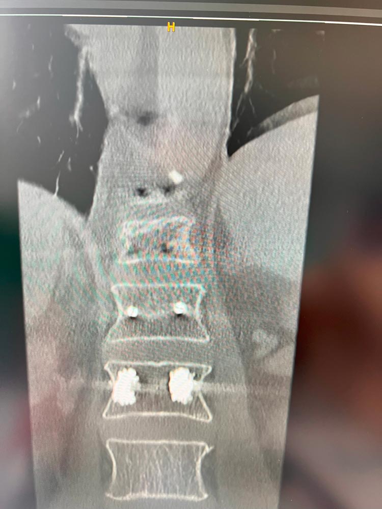
- Blood loss (intraoperative, total): 350 ml
- Duration of surgery: 210 min
- Post OP VAS: 2
- Neurological status: No problem
- Patient was discharged from the hospital after 7 days.

Published with the approval of Dr. Christoph Grimm


