E-Case report
Degenerative Disc Disease L2 – L5Dr. med. Mahmud Kadhem
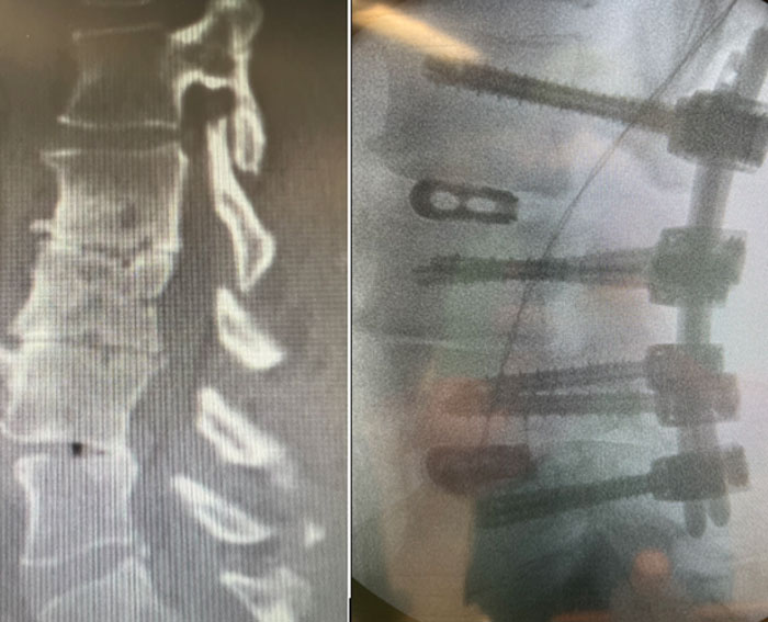
Pre OP

Clinical Case – Degenerative Disc Disease L2 – L5
Mahmud Kadhem, MD
Neurosurgeon
Klinik Kitzinger Land
Department of Neurosurgery
Würzburg-Kitzingen, Germany


Patient Information
Female 48 years old, obese patient was diagnosed with a degenerative disc disease (DDD) in the lower lumbar area.
- Instabilty, abrasion and pain from the facet joints in levels L2/3, L3/4, L4/5
- Osteochondrose, and coronal disbalance
Bone quality: Good
Back pain: Pre OP VAS: 5-6
Neurological status: OK
Revision surgery


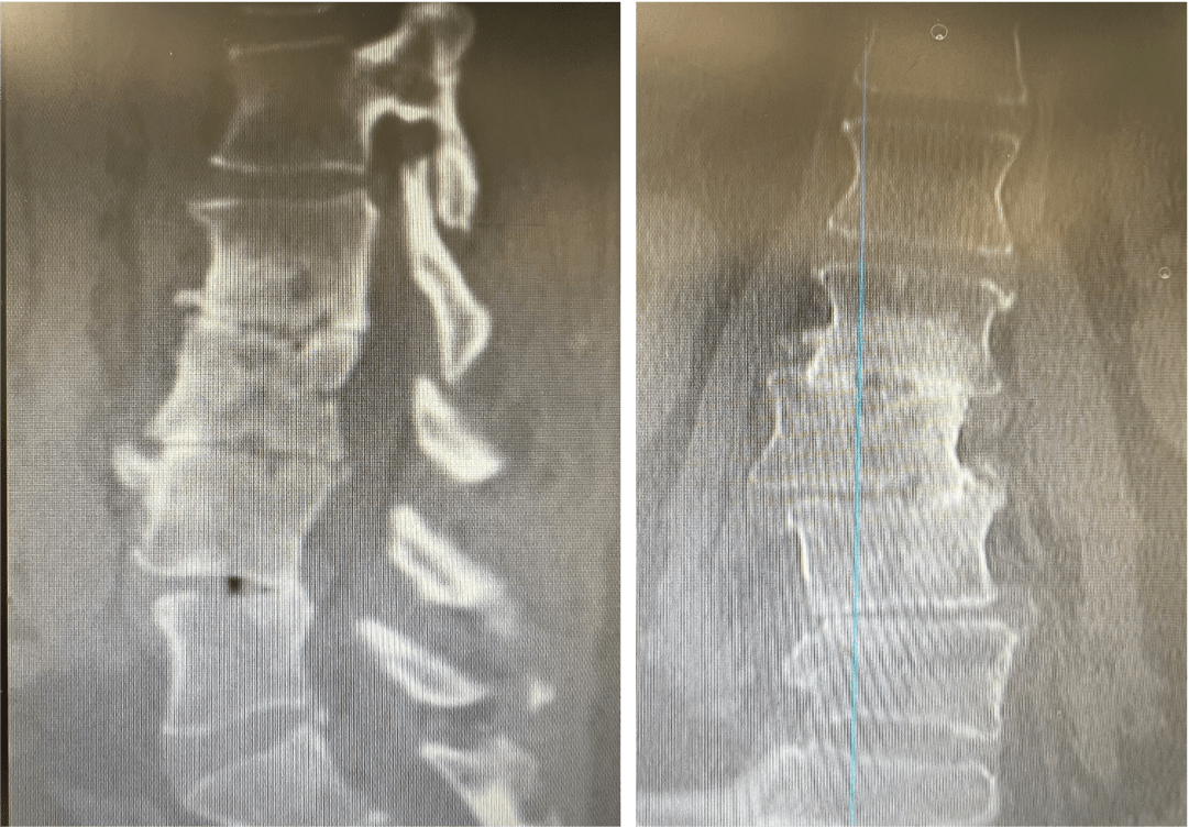
Pre OP CTs,
Sagittal & Coronal
The coronal view confirmed the disbalanced spine

The surgery was planned using the Neo Pedicle Screw System™ L2 – L5, and the Neo Cage System™ in levels L2/3 and L4/5


Intra OP

Intra OP
The patient was placed in prone position, intubated, general anesthesia ITN & TIVA.
The percutaneously midline cut was followed by decompression and preparation of the disc space.
Four Neo Medical Cage System™ PLIF cages were implanted from the left side.
In level L4/5: 2 x Cage anatomical/straight 22mm, 9-10mm, 8°
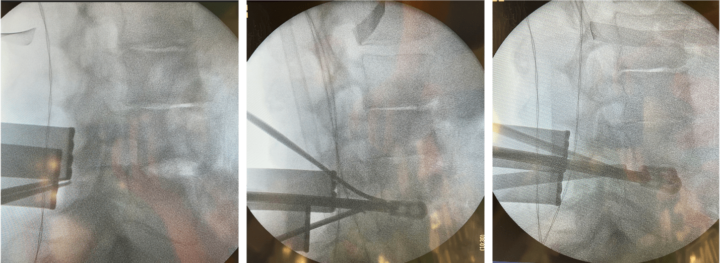


The intervertebral L3/4 level was already bony fused.
In level L2/3: 2 x Cage anatomical/straight 22mm, 7-8mm, 8° were implanted.
Bone graft: Nuvasive Attrax Putty
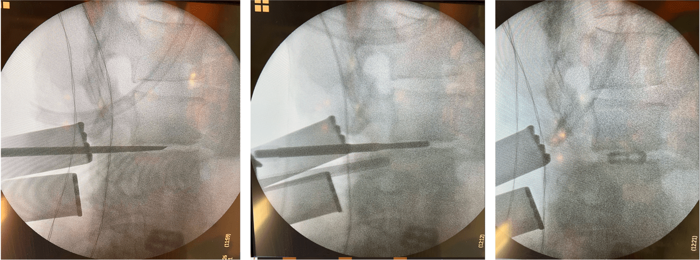


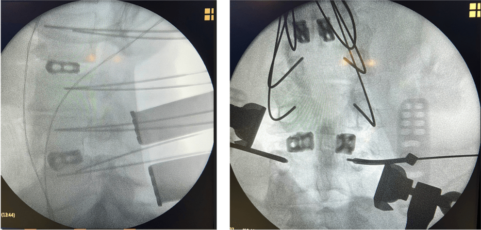
Neo pedicle screw system, polyaxial screws, was used to fuse segments L2 – L5
L2: 2 x Ø6.0×50 mm
L3: 2 x Ø6.0×50 mm
L4: 2 x Ø7.0×45 mm
L5: 2 x Ø7.0×45 mm
2 x 70 mm straight titanium rods
Duration of surgery: 450 min
Total blood loss: 300 ml
K-Wire – placement (lateral) L2 – L5

Post OP
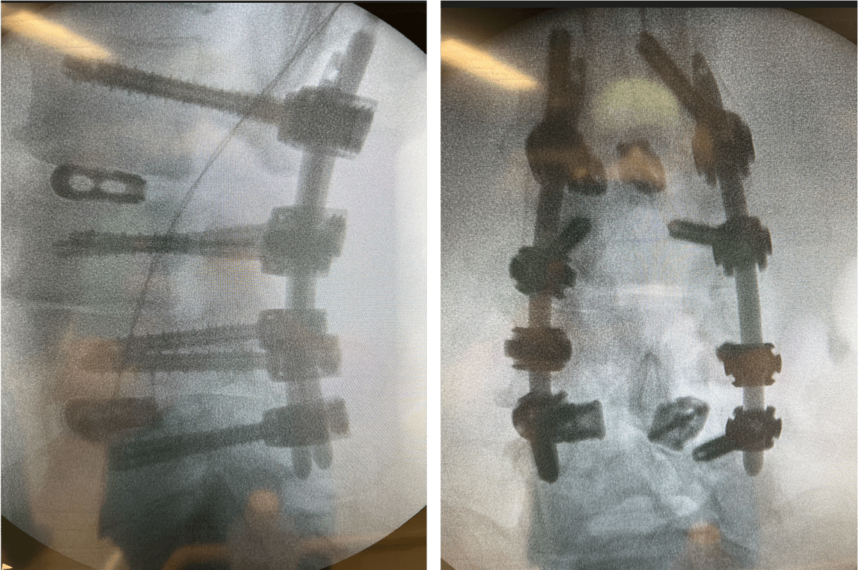
Post OP Radiographs, sagittal and frontal views
The patient is discharged from the hospital after 8 days
Pain VAS:2 at discharge

Published with the approval of Dr. Mahmud Kadhem, MD


