E-Case report
Trauma fractures T10 & T11
Posterior fixation T9 – L1 Tino Beylich, MD

Pre OP

Clinical Case – Trauma fractures T10 & T11
Posterior fixation T9 – L1
Neo ADVISE™ Trauma module
Tino Beylich, Chief of trauma department
Orthopaedic Trauma Surgeon
Klinikum Bad Salzungen GmbH
Clinic for trauma surgery & orthopaedics
Bad Salzungen
Germany


Patient Information:
Young male patient, 36-year-old, diagnosed with painful traumatic fractures in T10 and T11. No neurological deficits and good bone quality.
In addition, the patient suffered from serial rib fractures and pulmonary contusion. Alcohol and nicotine abuse is present.
Planned Surgery:
- General anesthesia
- Percutaneous MIS surgery, lateral approach
- Fracture reduction T11 – mono-axial screws in T10 & T12
- Posterior fixation T9 – L1
- Neo ADVISE™ trauma module to be used

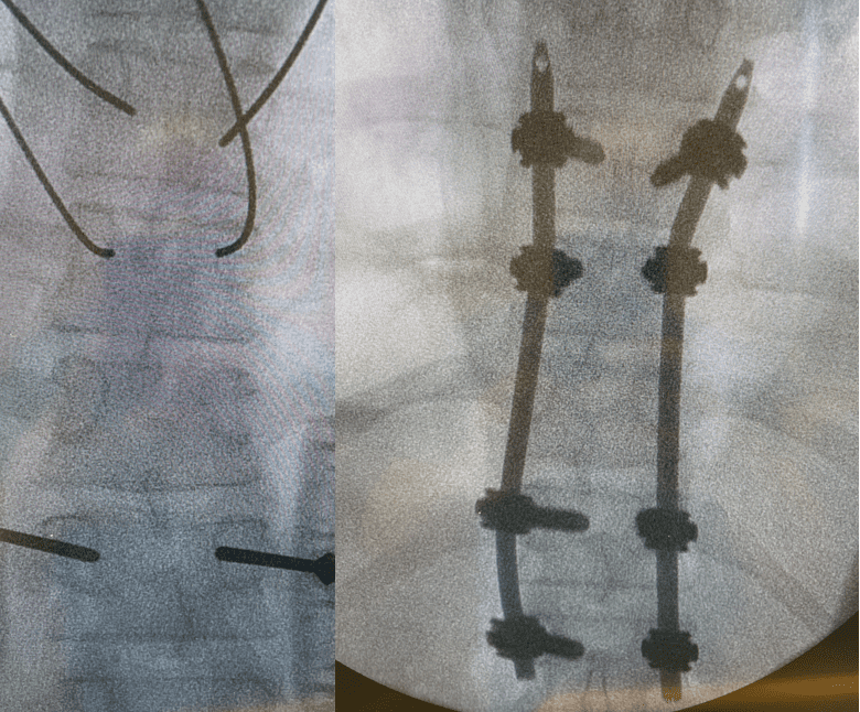
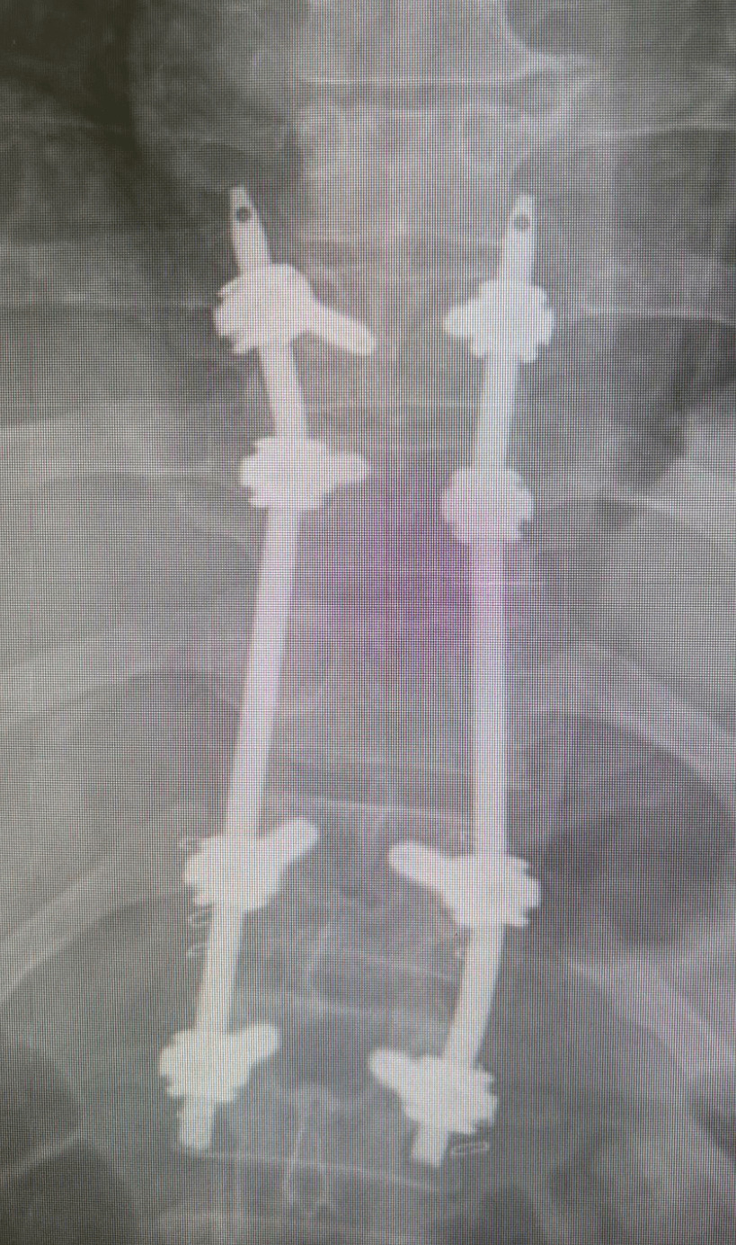
Pre OP radiograph, frontal view


Intra OP
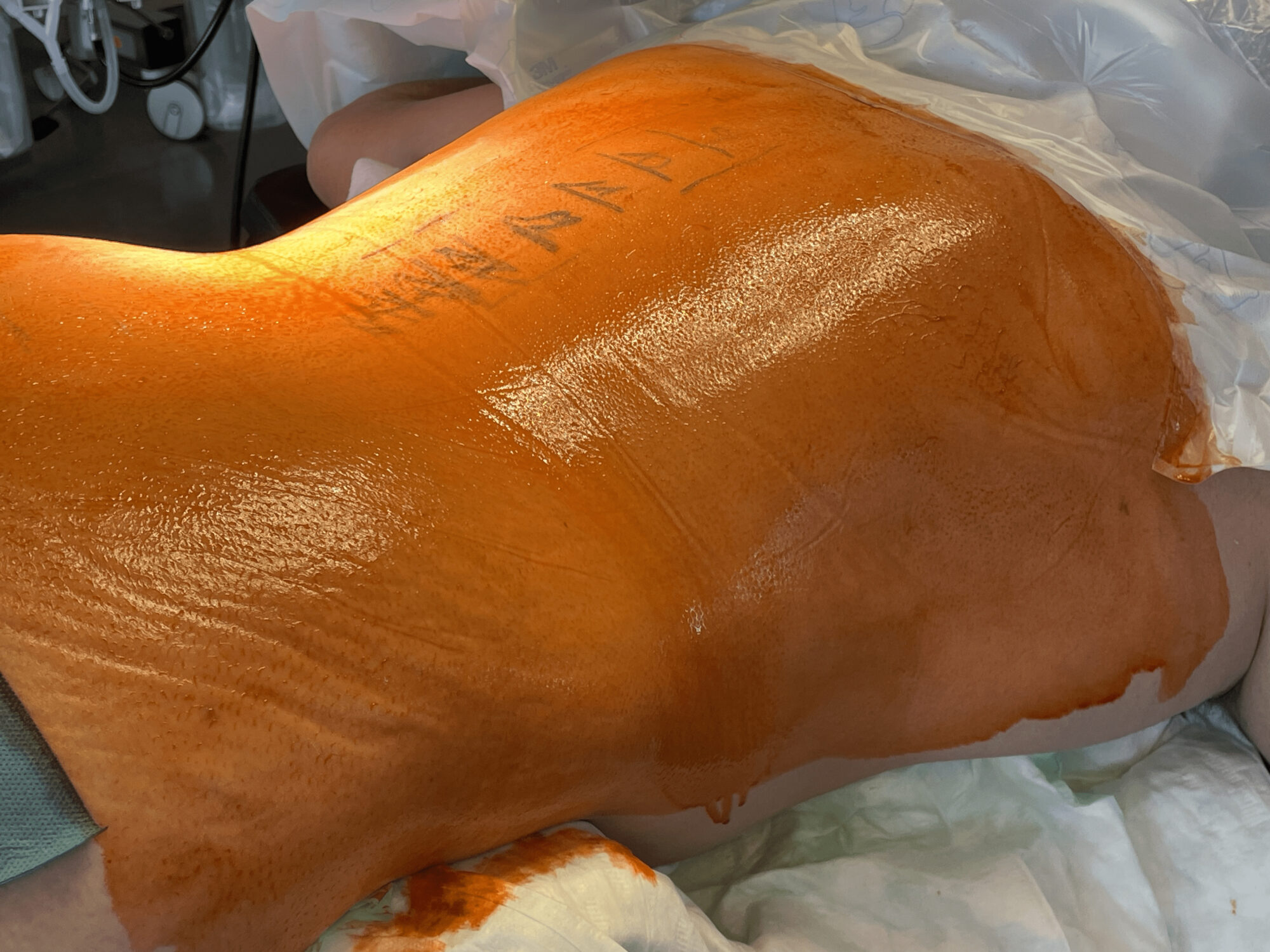
Patient was placed in prone position, good correction achieved with ligamentotaxis.
Percutaneous MIS surgery, lateral approach from cranial to caudal, starting at T9 level.
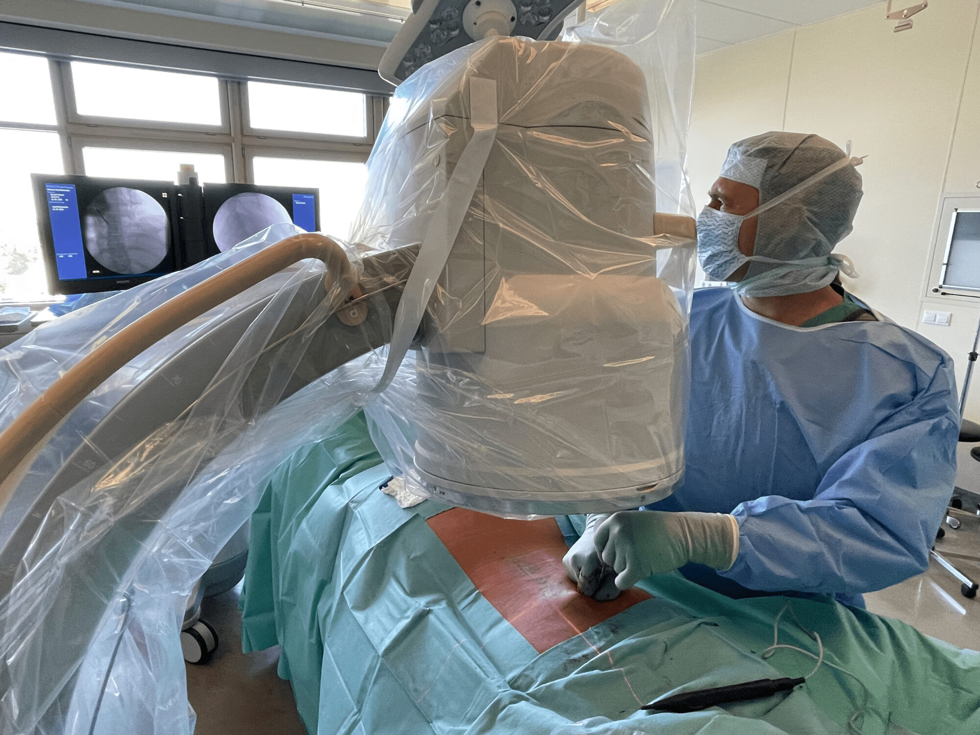
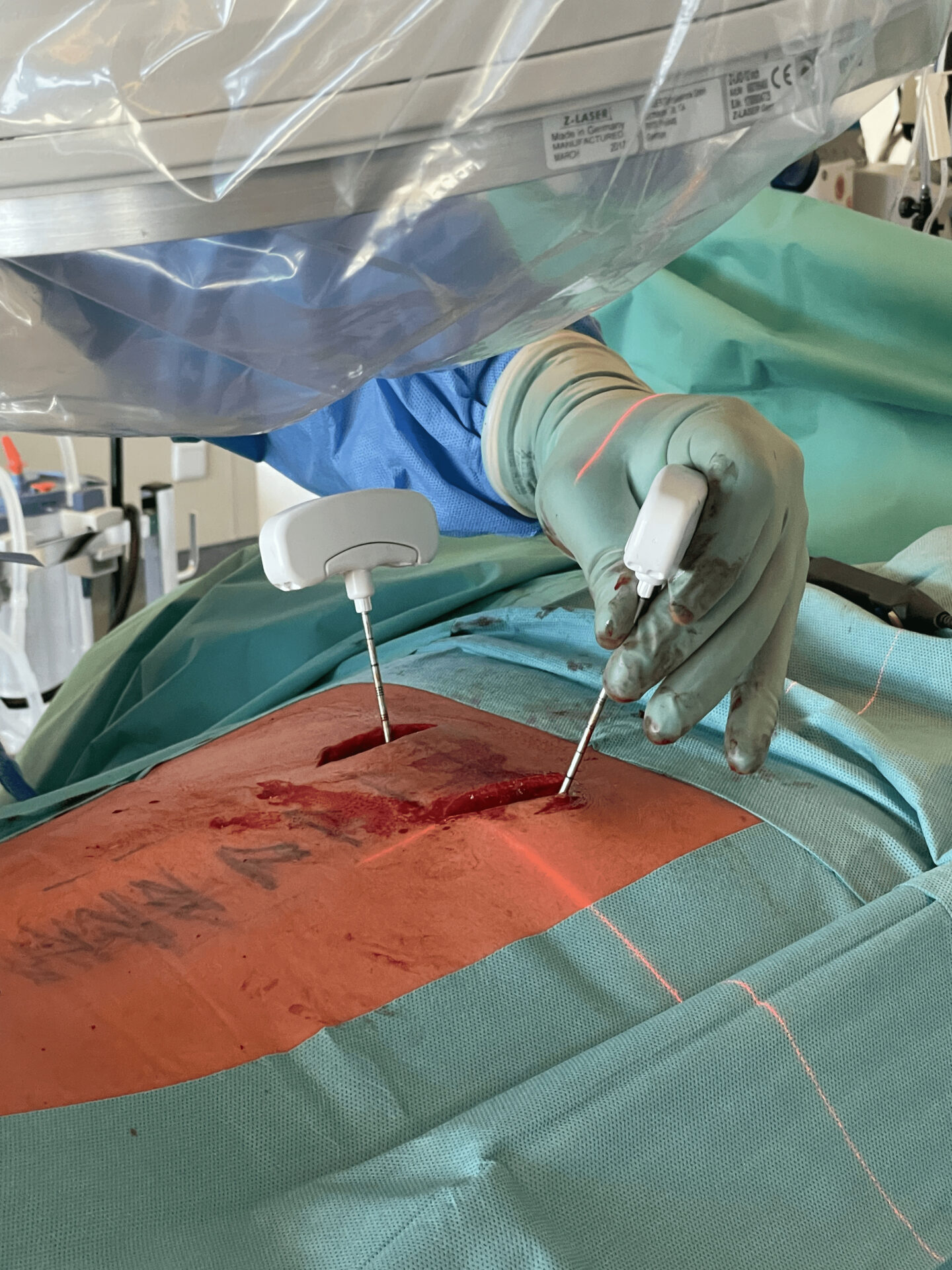
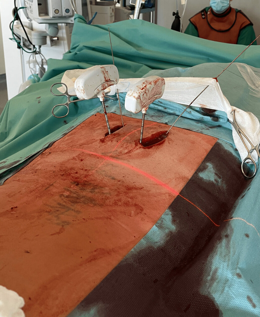

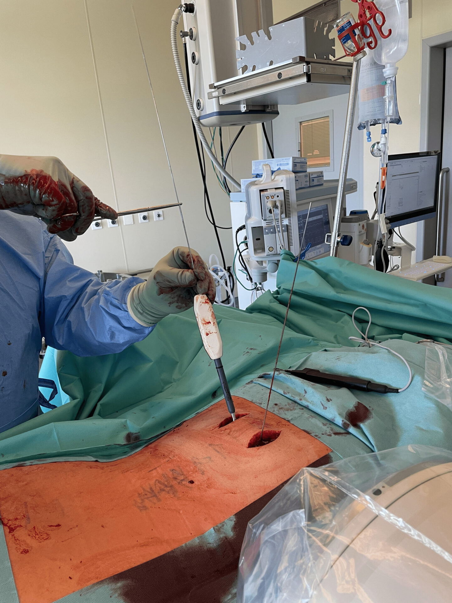
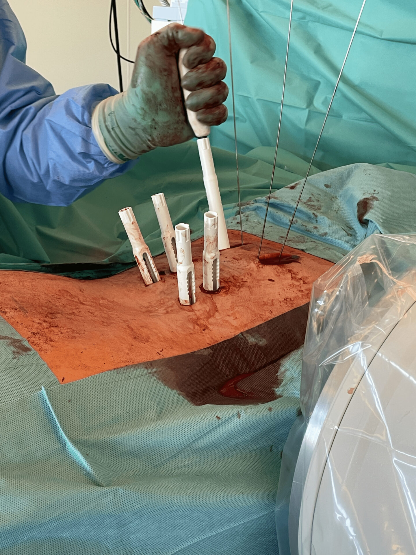
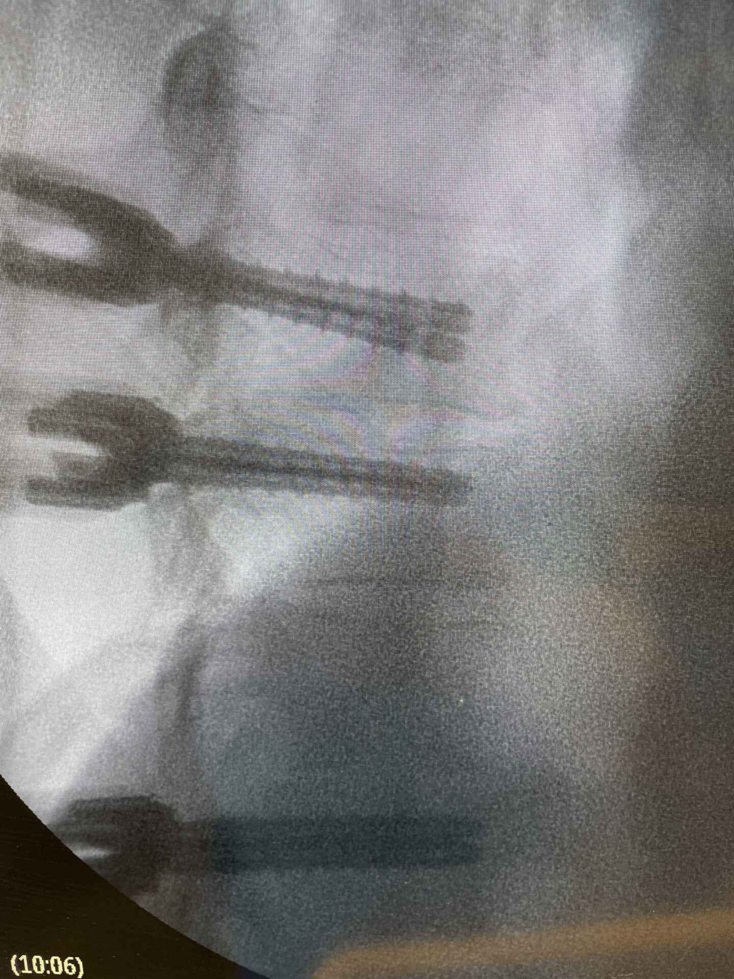
Inserting the K-wires at each level
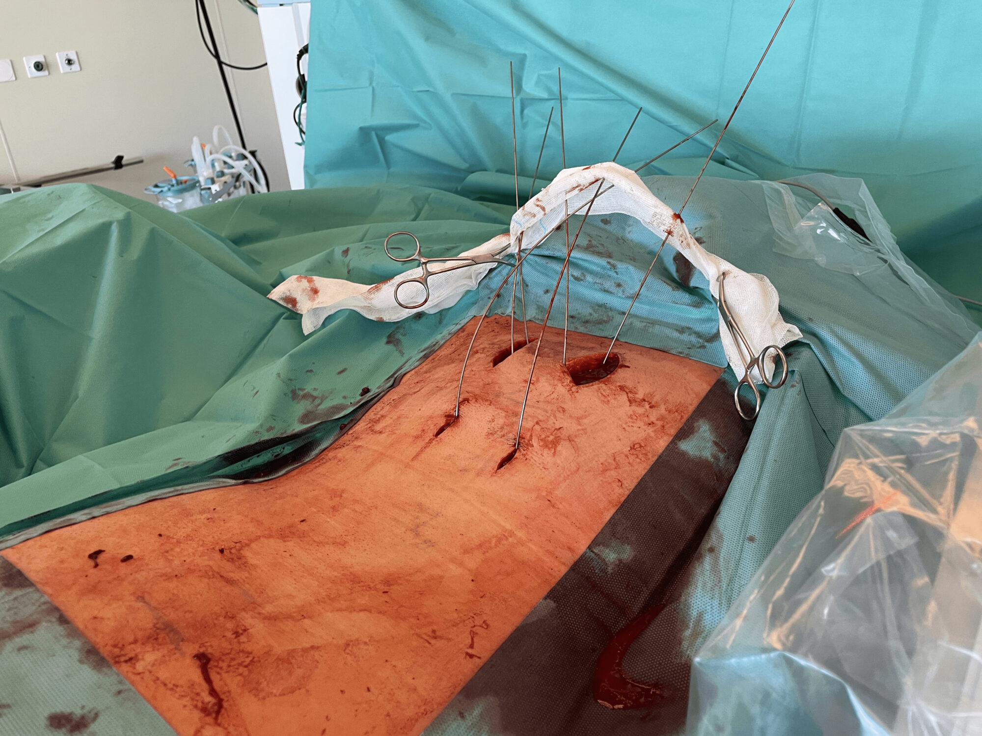
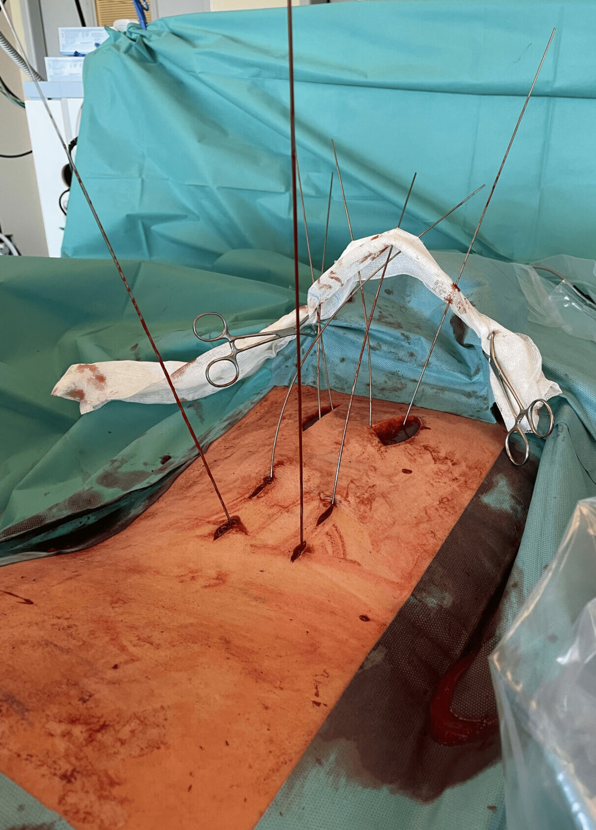
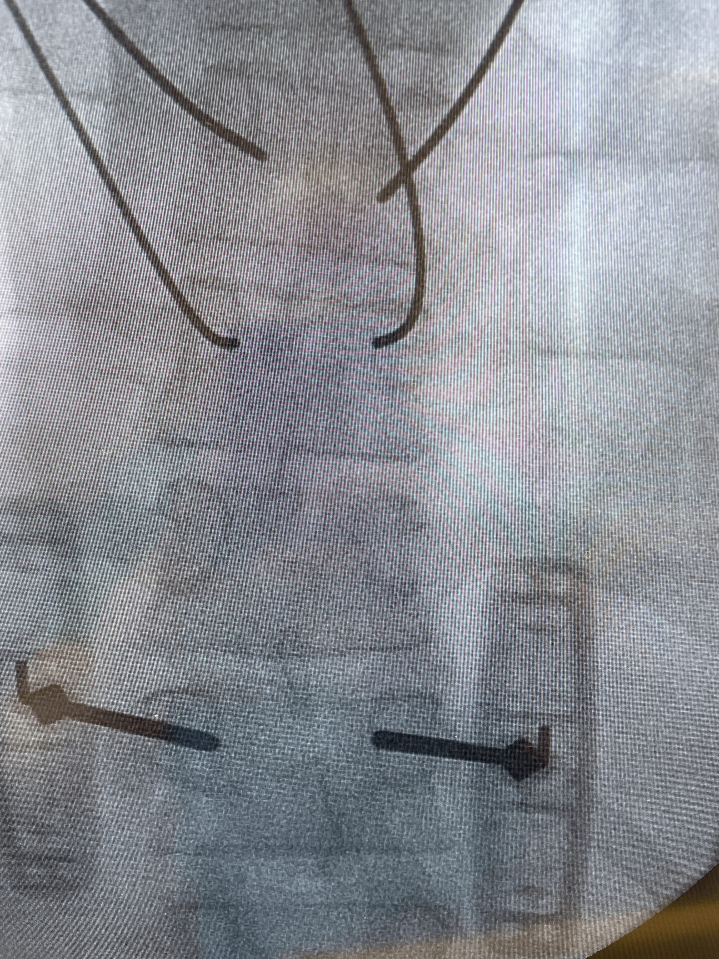
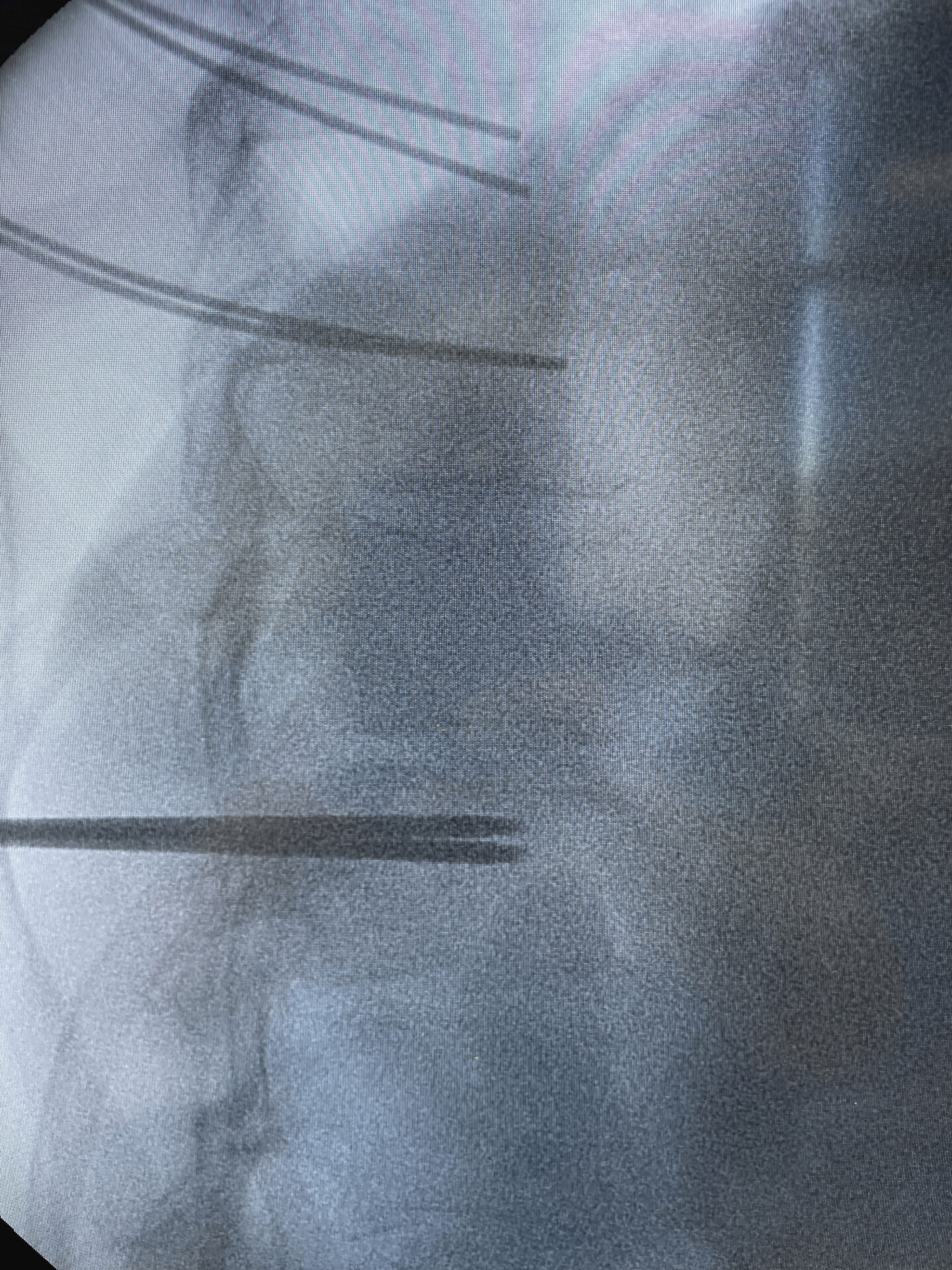

Pedicle screws insertion
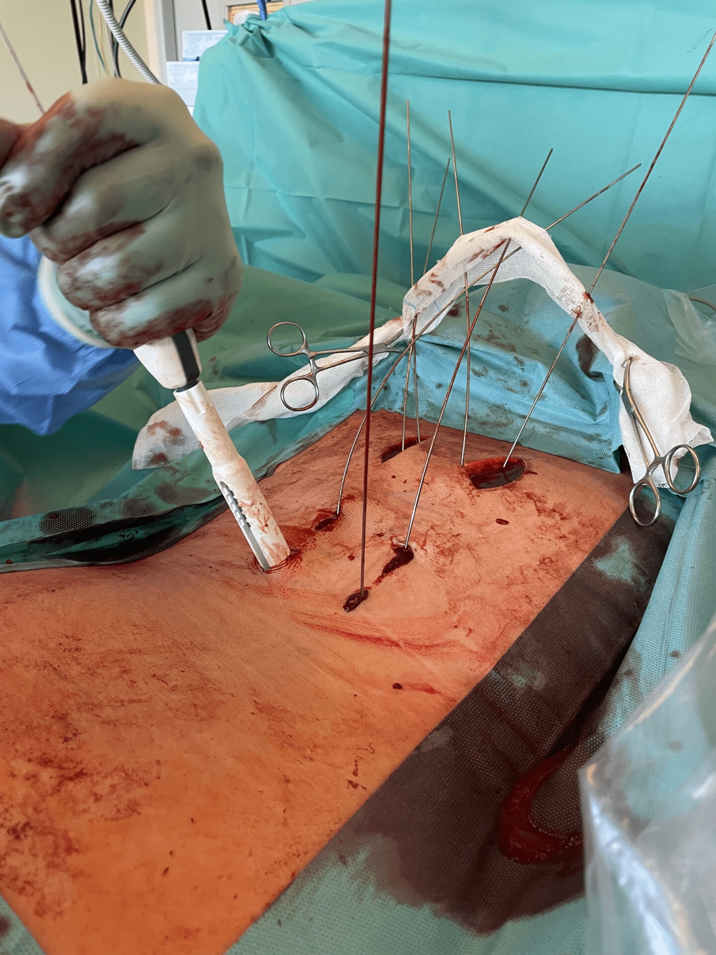
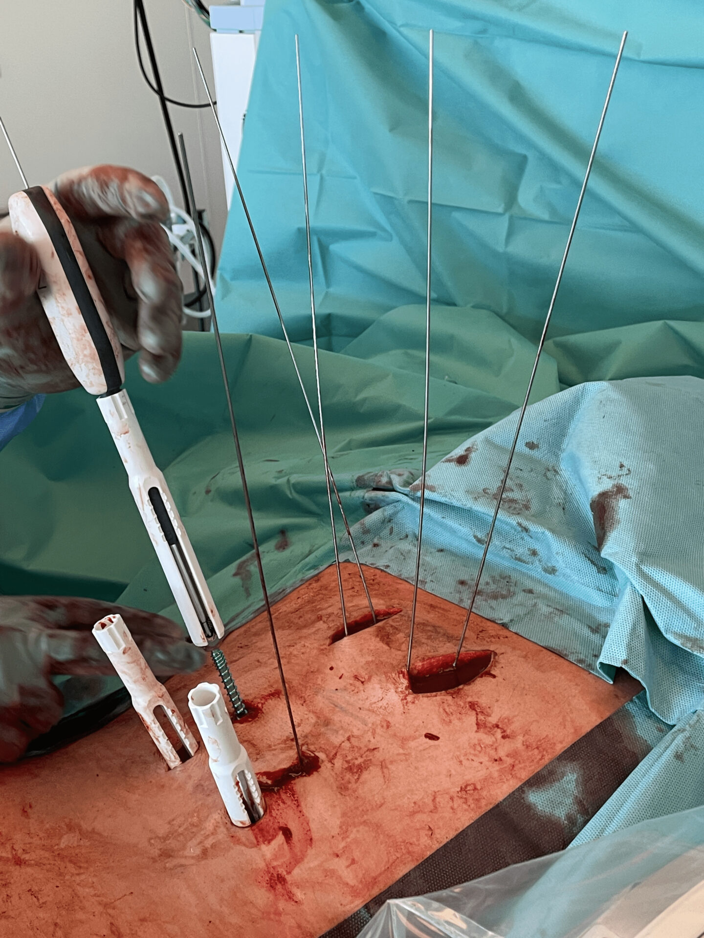

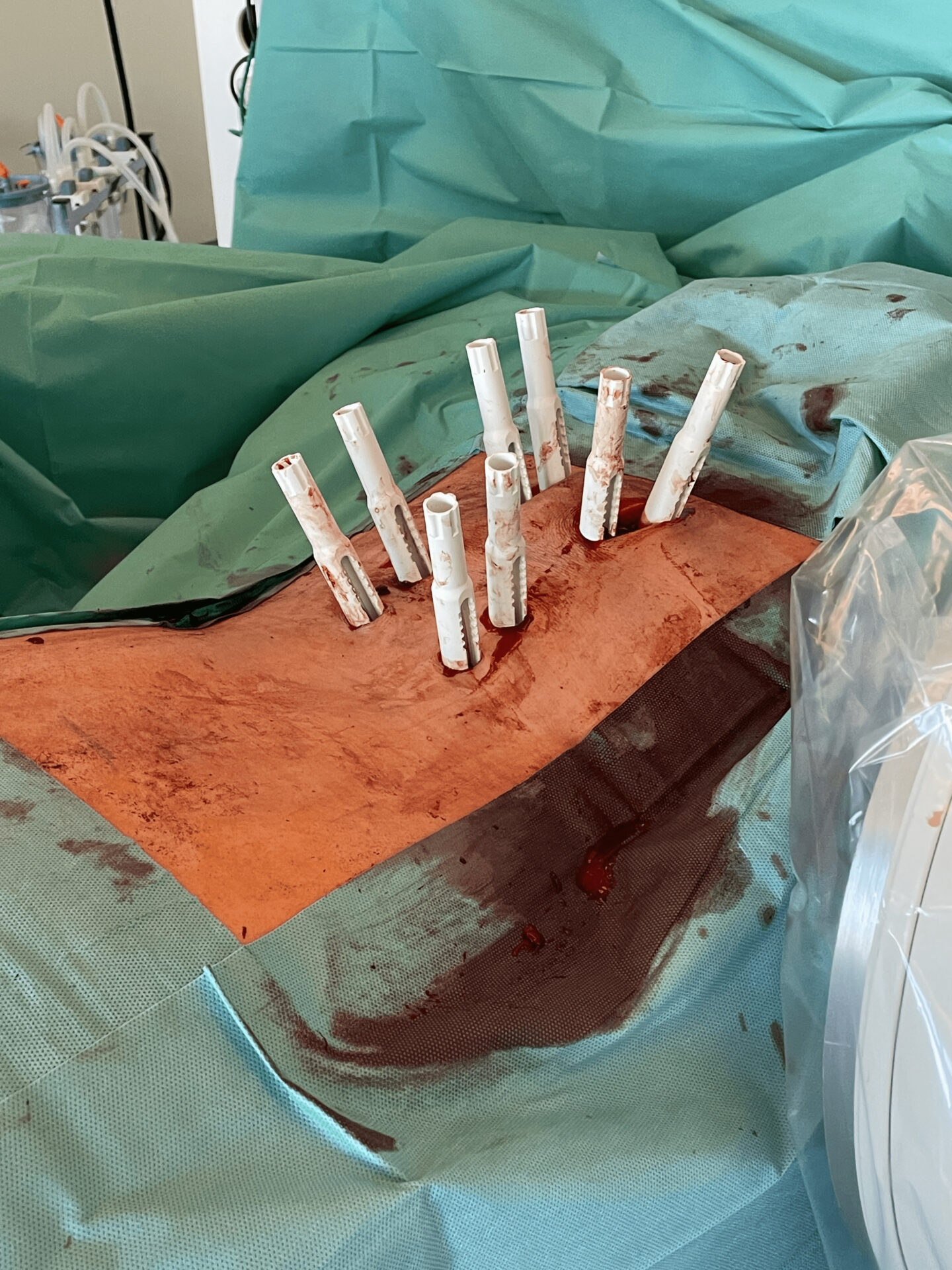
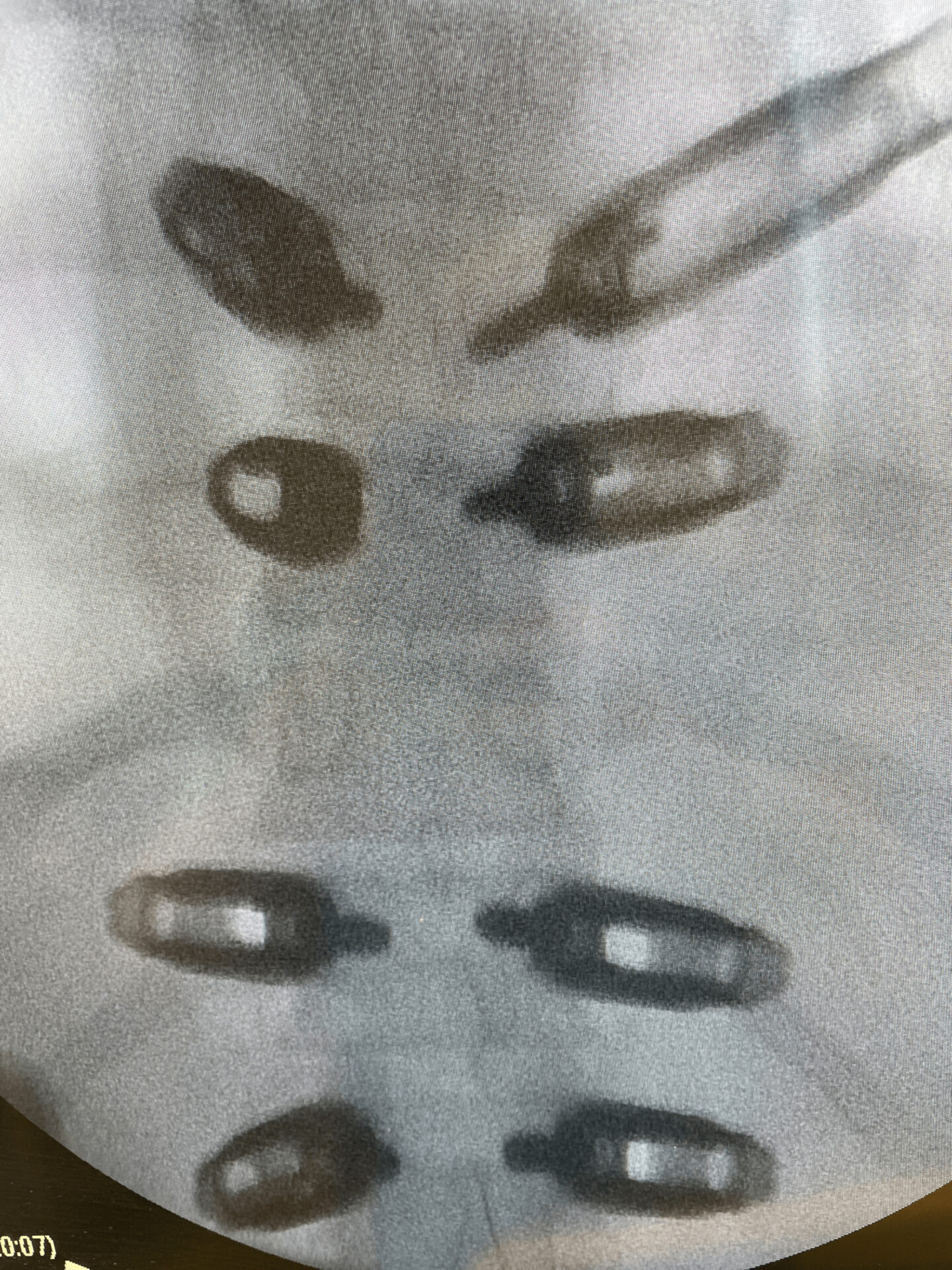
Posterior fixation with Neo Pedicle Screw System™
Totally 8 pedicle screws placed over the levels T9 – L1
- T9 2 x Ø6.0x45mm polyaxial
- T10 2 x Ø6.0x45mm monoaxial
- T12 2 x Ø7.0x50mm monoaxial
- L1 2 x Ø7.0x50mm polyaxial
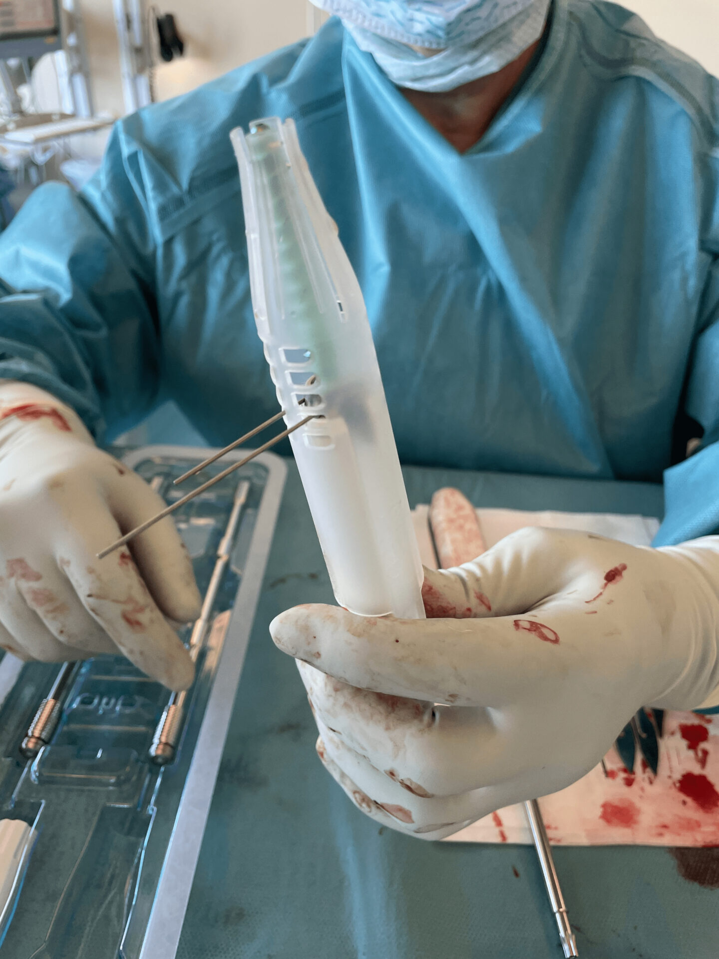
The surgeon decided to use the monoaxial capability of the Neo pedicle screws in two of the levels next to the fractured T11 vertebra.
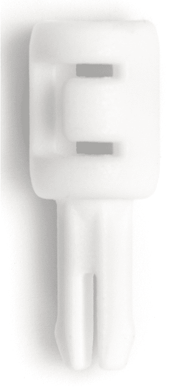
The clip, available in all Neo pedicle screw kits, can be used and inserted to lock the screw head in a monoaxial position.


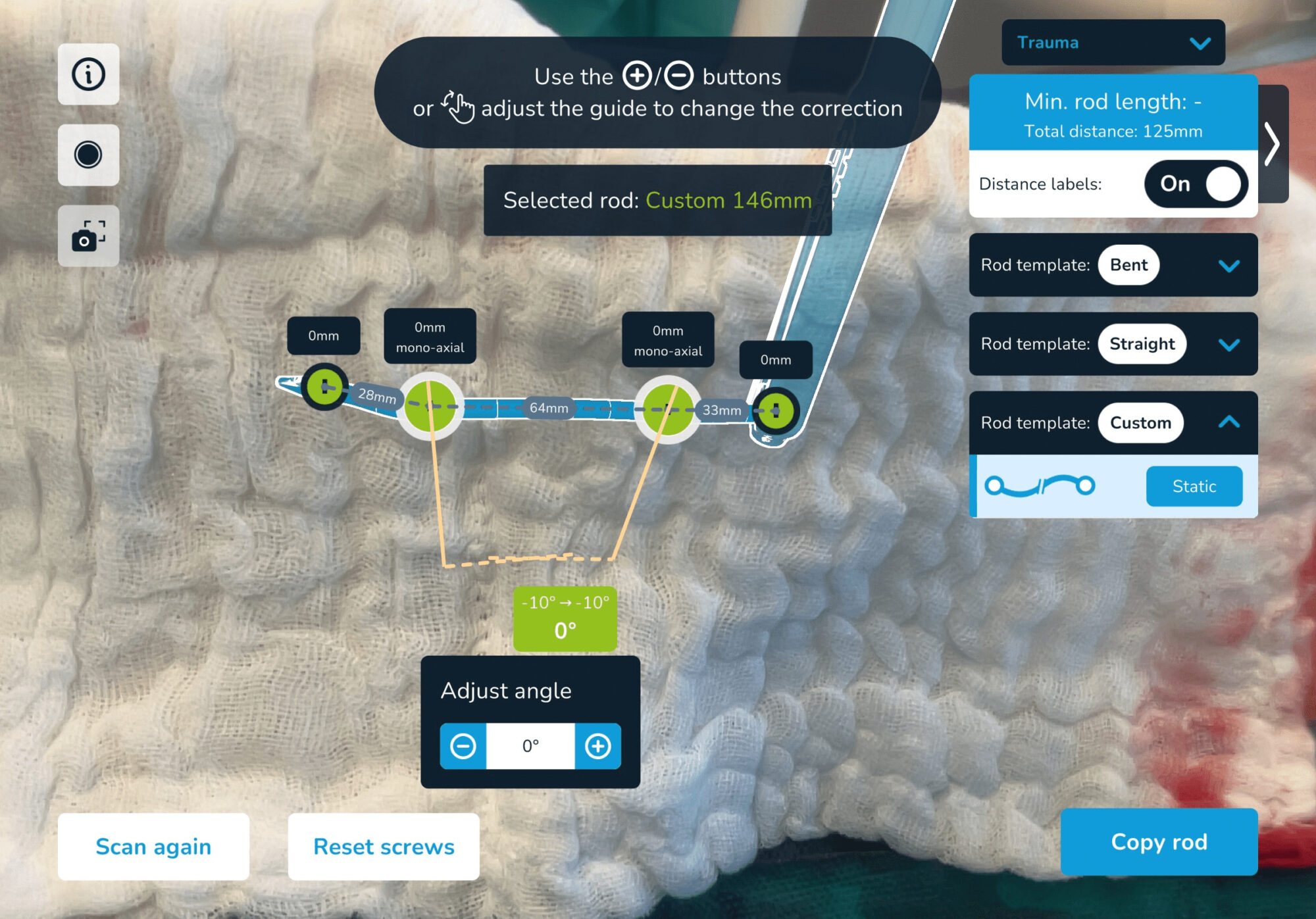
Usage of Neo ADVISE™ trauma module
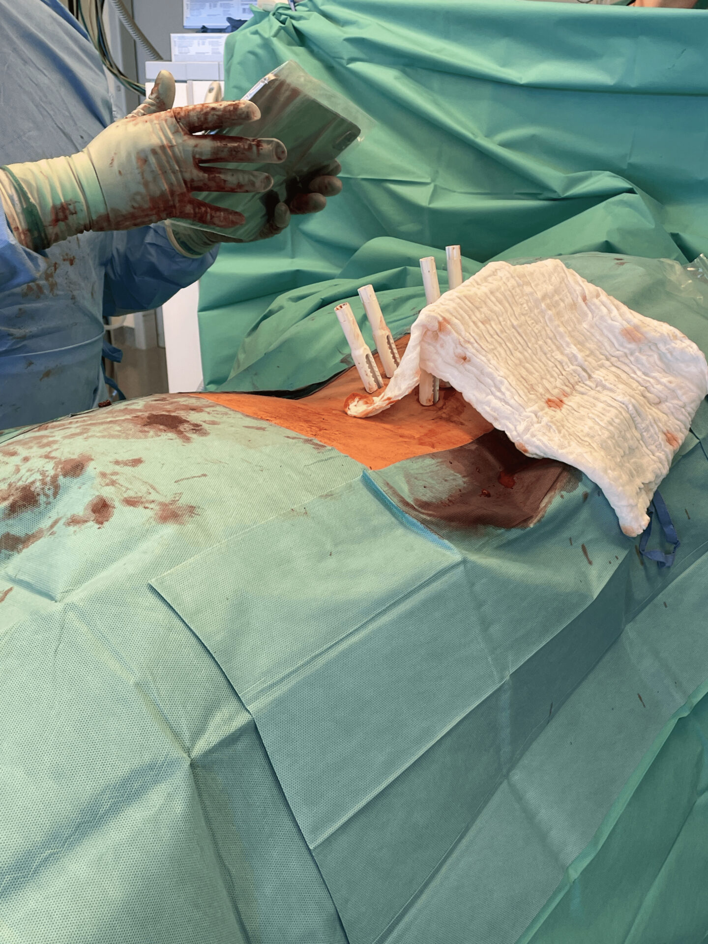
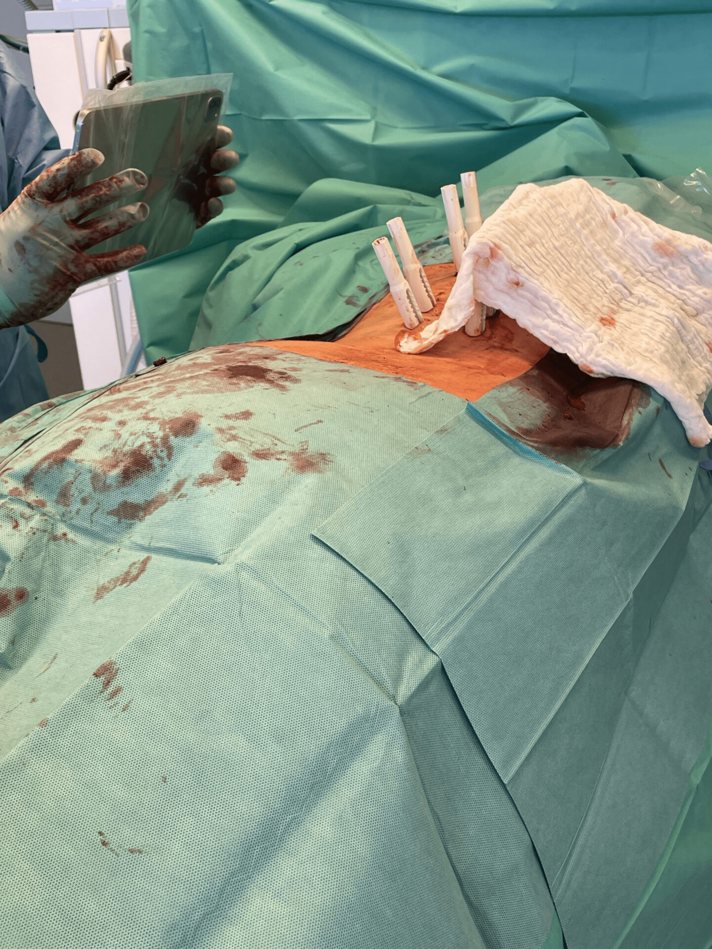
ADVISE™ scene analysis and 3D mapping of the guide towers at the patient´s right side.


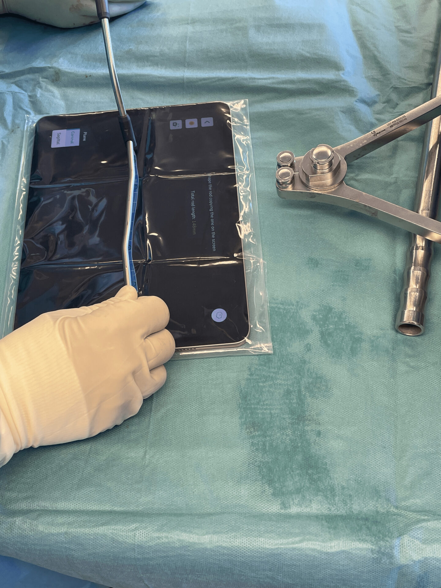
Results from using the Neo ADVISE™ trauma module in both sides and after the targetted angle correction has been achieved. Visualizations of the rods in both the sagittal and the coronal planes in the iPad screen.
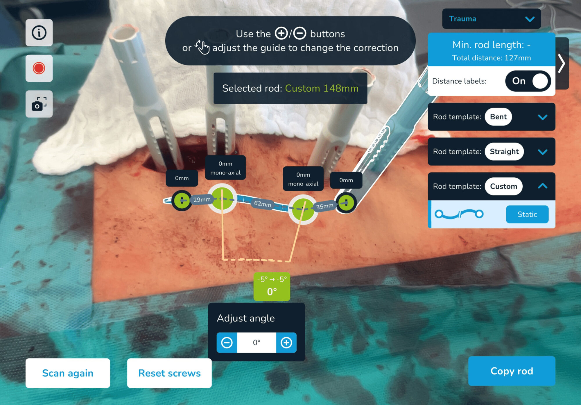
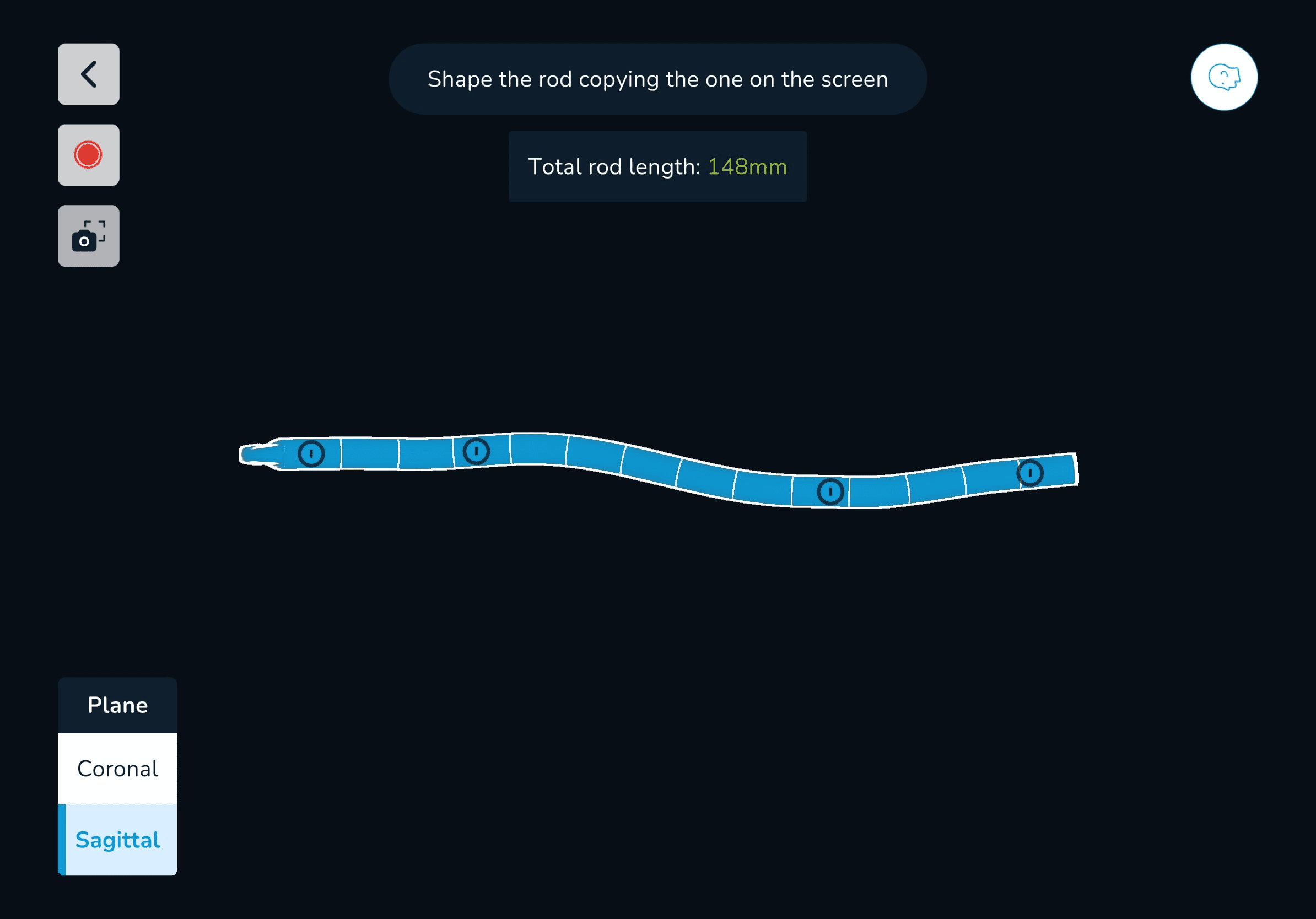
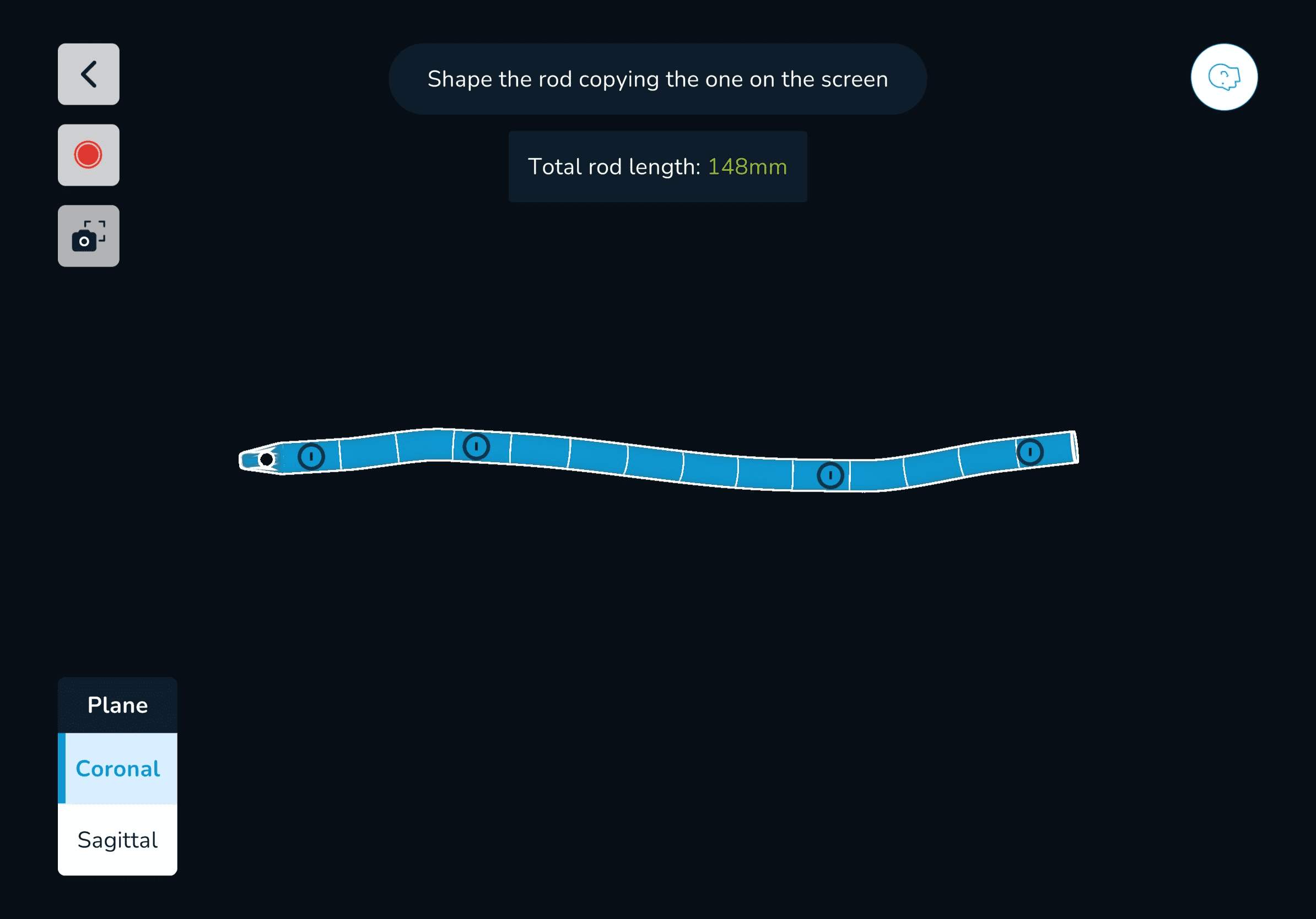
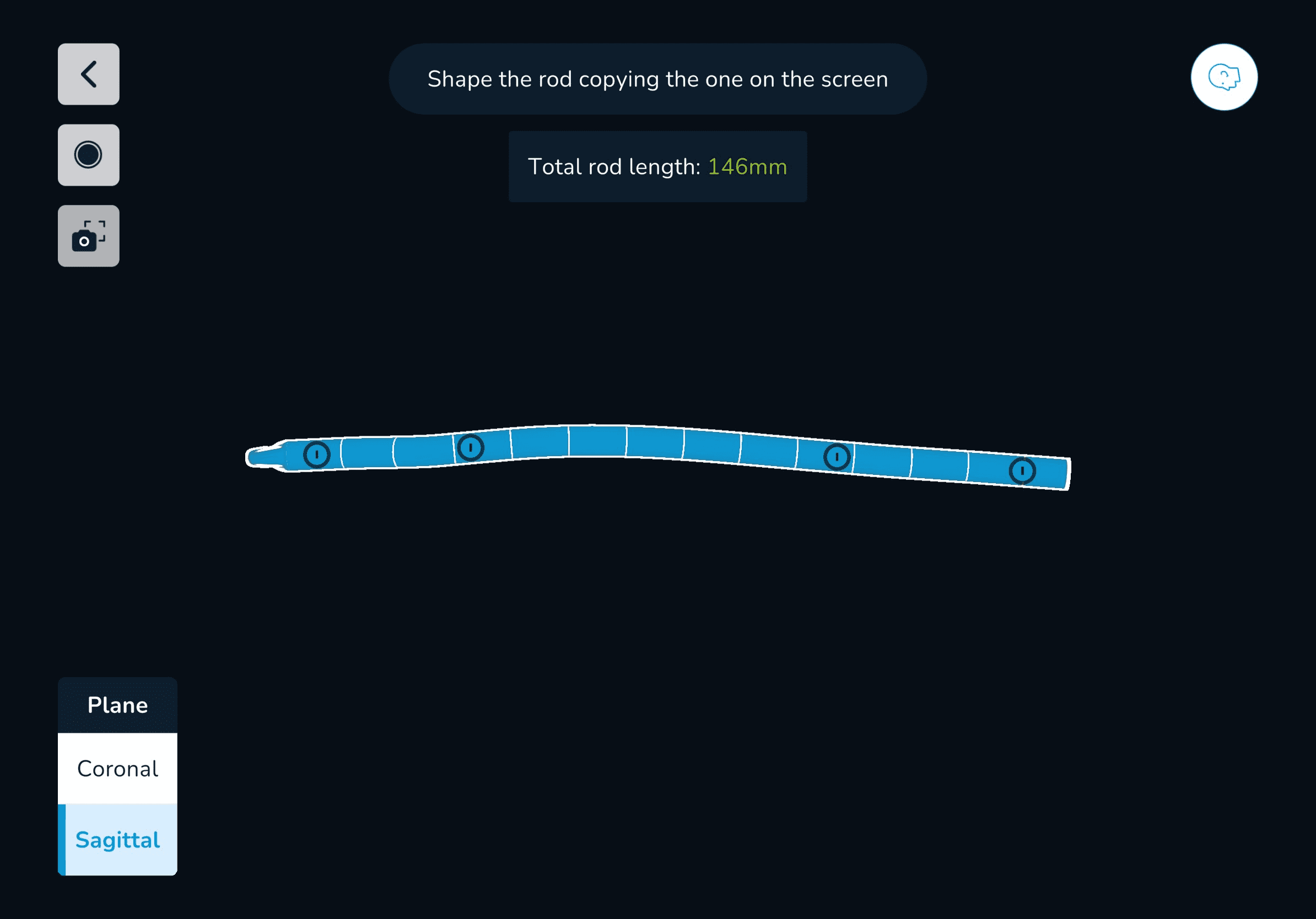
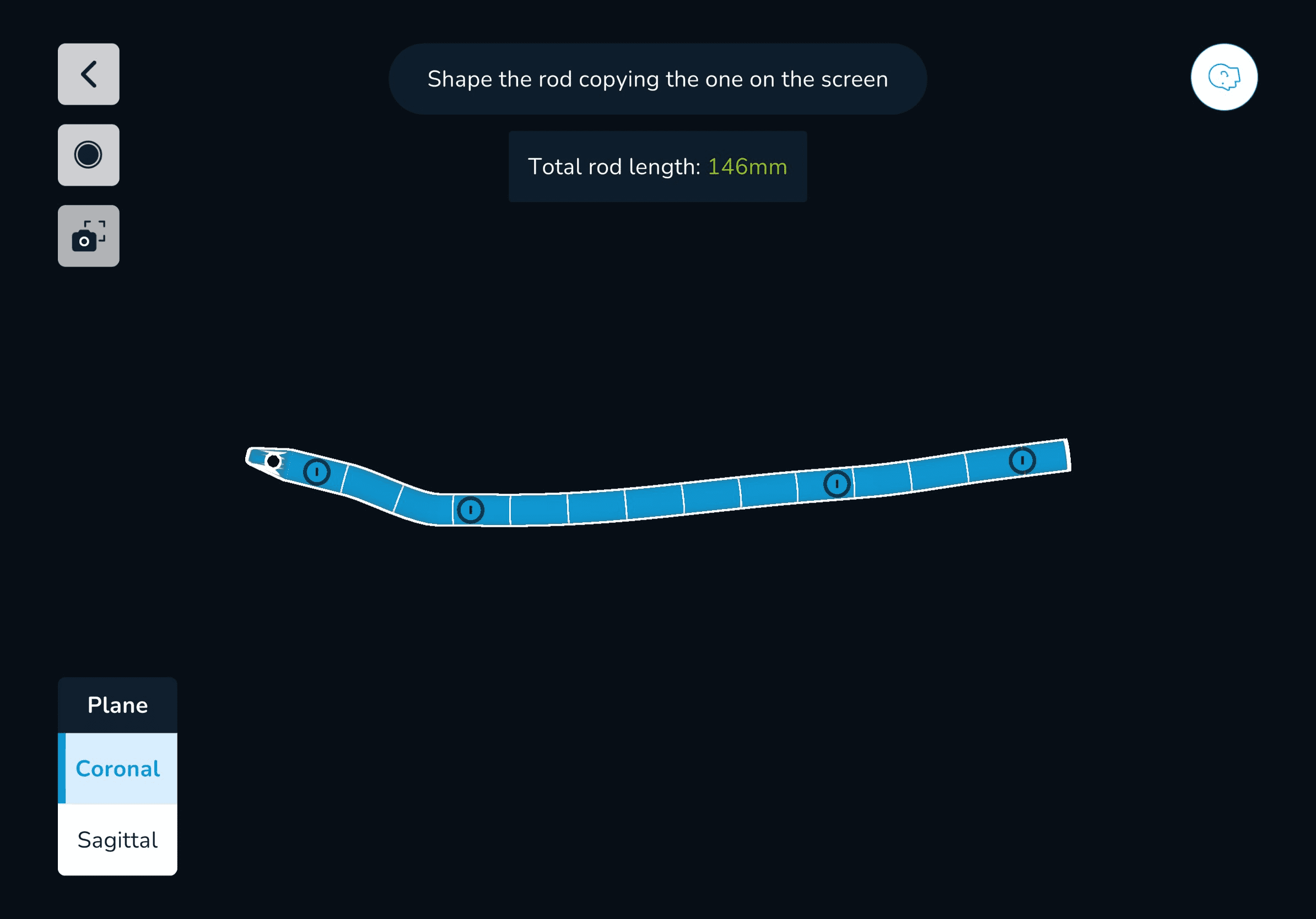
Rod selection: 2 x Titanium rods 160mm straight straight and shortened to 150mm


The video is showing all steps using Neo ADVISE™ 3D scanning trauma module


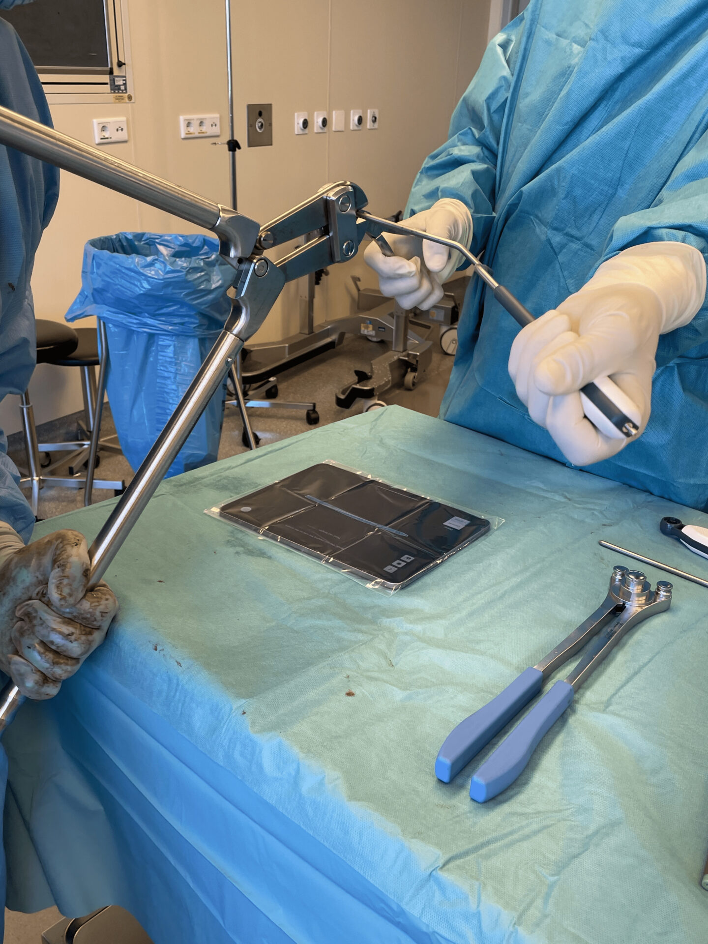
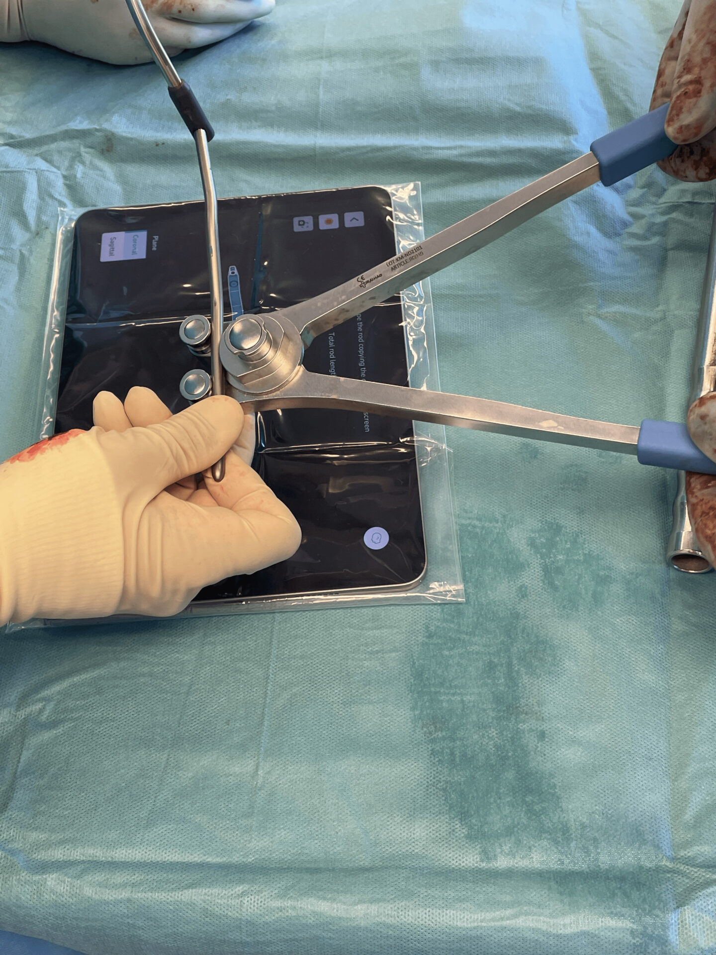
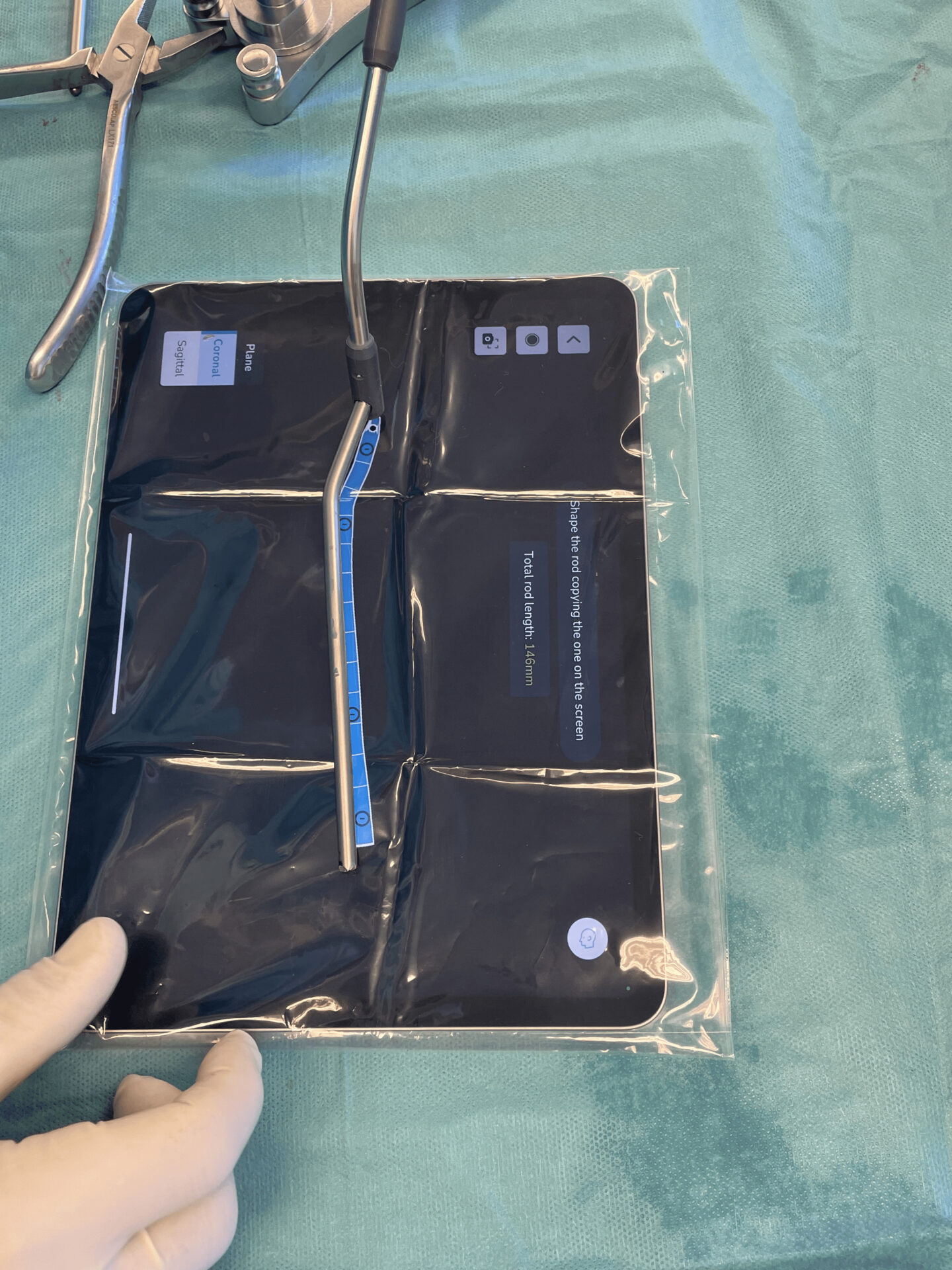
Rod bending: using the rod visualization in the iPad screen as template for cutting and contouring the rods in both the sagittal and the coronal planes.


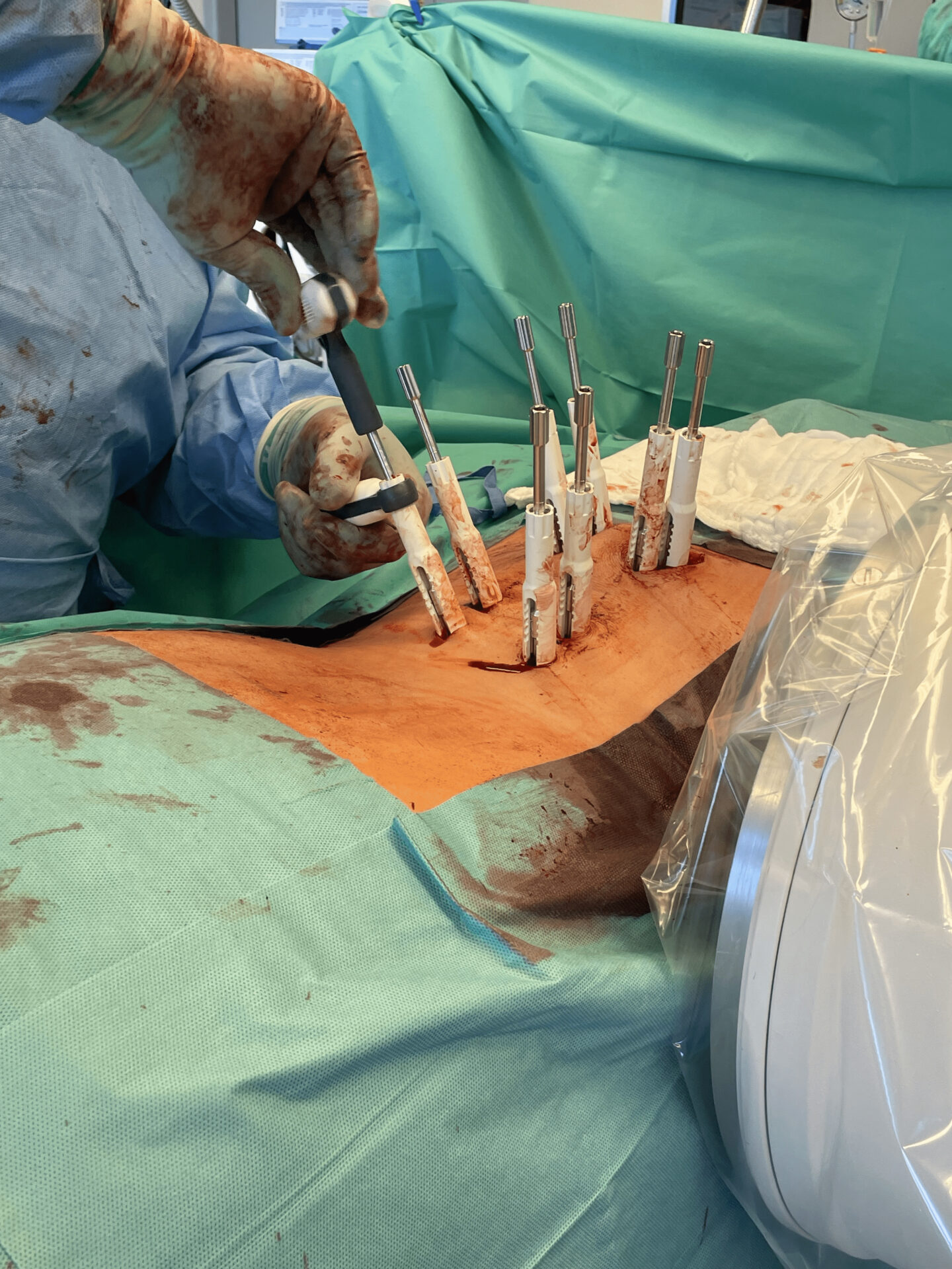
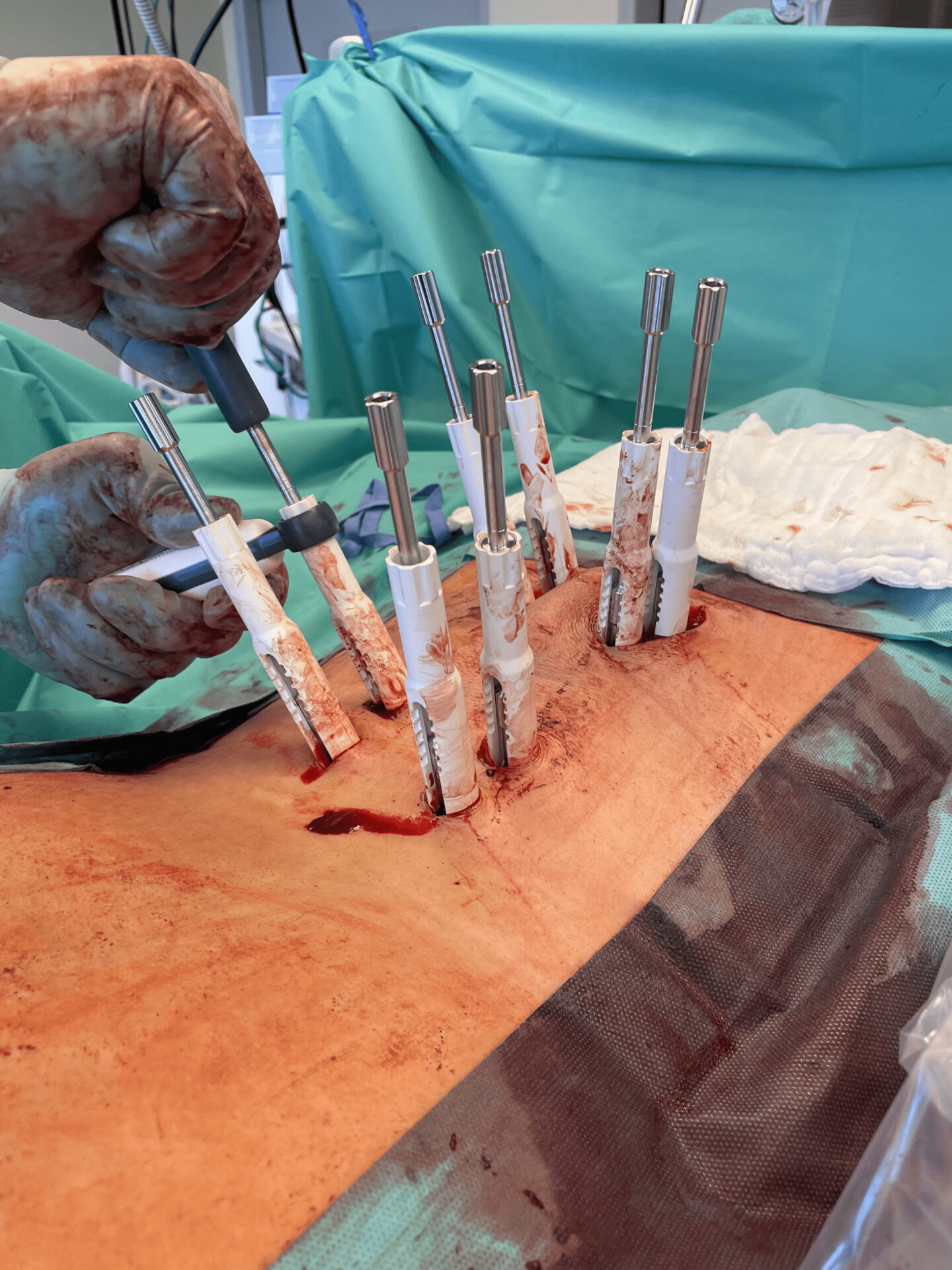
Rod insertion, from cranial to caudal, and pre-tightening of the most caudal screws using the torque limiter in left and right side.
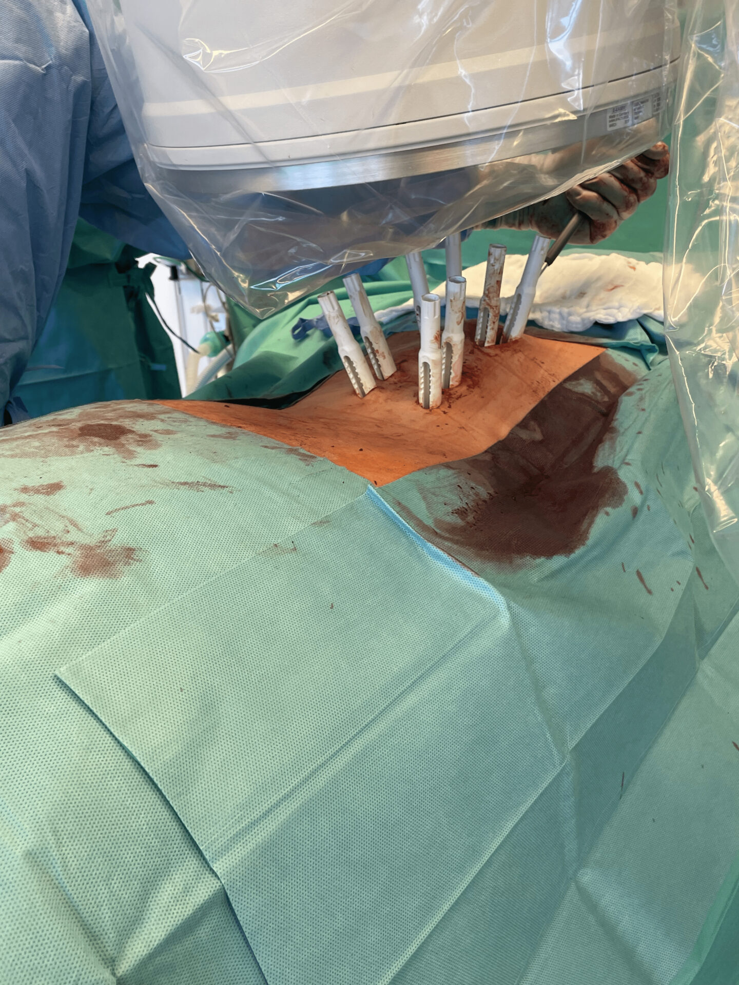
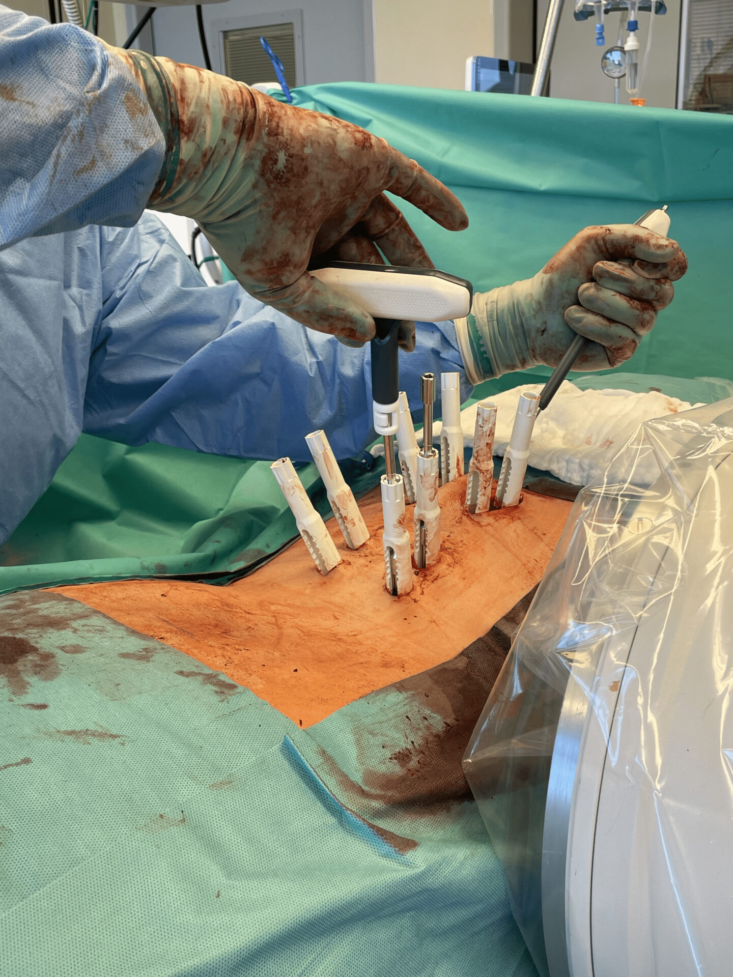
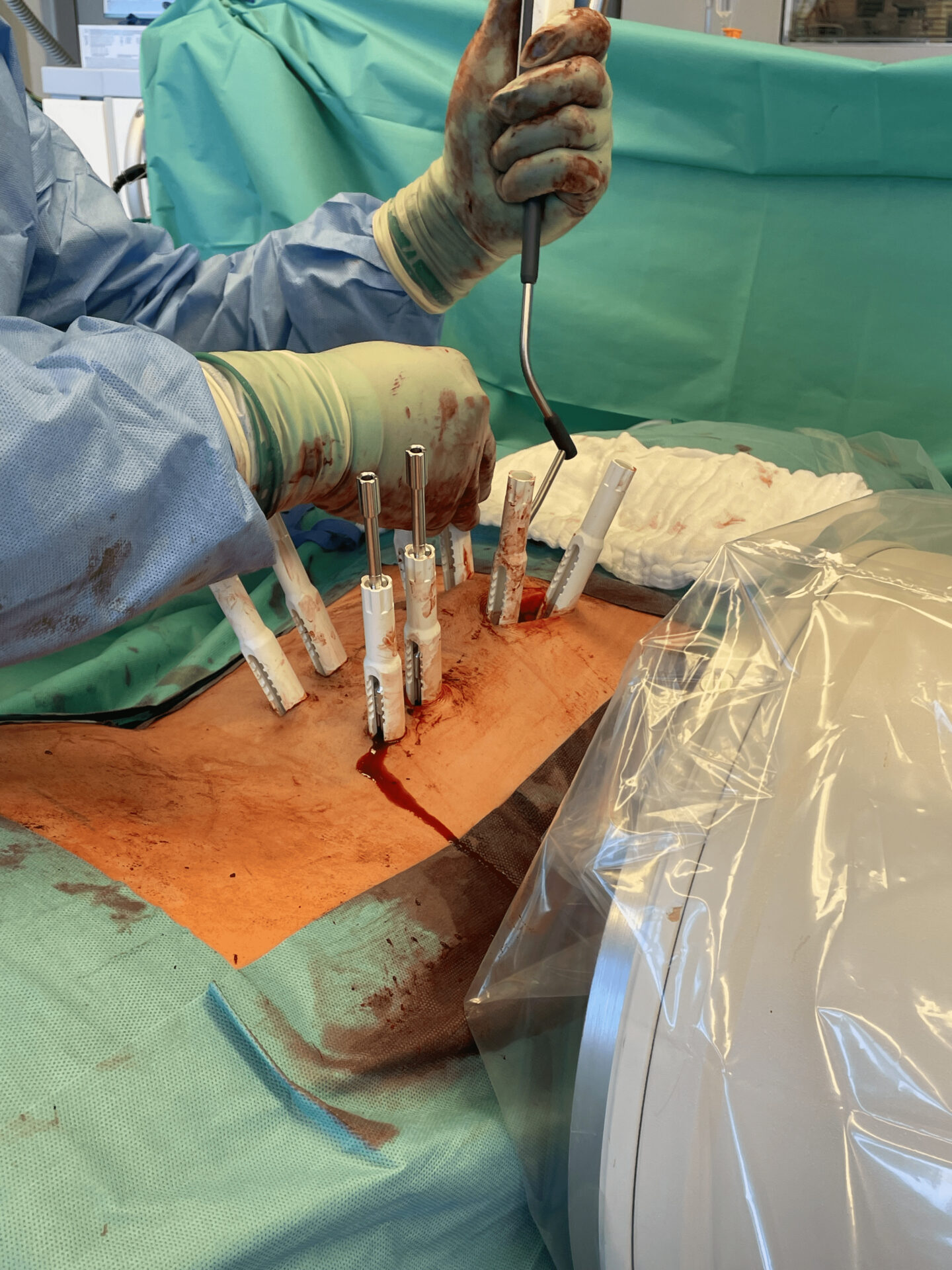
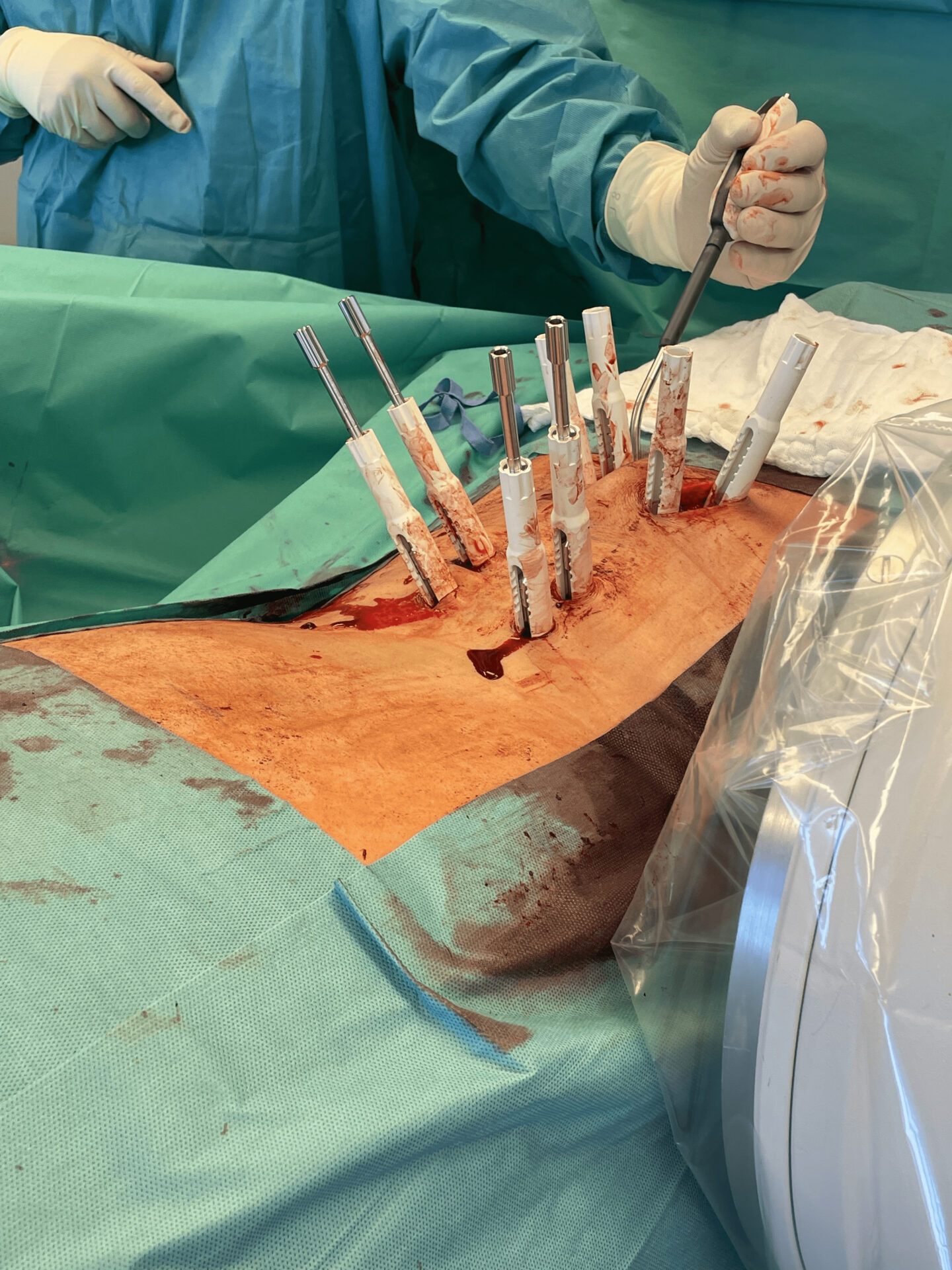
The torque limiter is placed between the T-handle and the set screw driver. Pre-tightening is achieved when the torque limiter is reached with an audible ≪ click – click ≫


Final set screw tightening from caudal to cranial


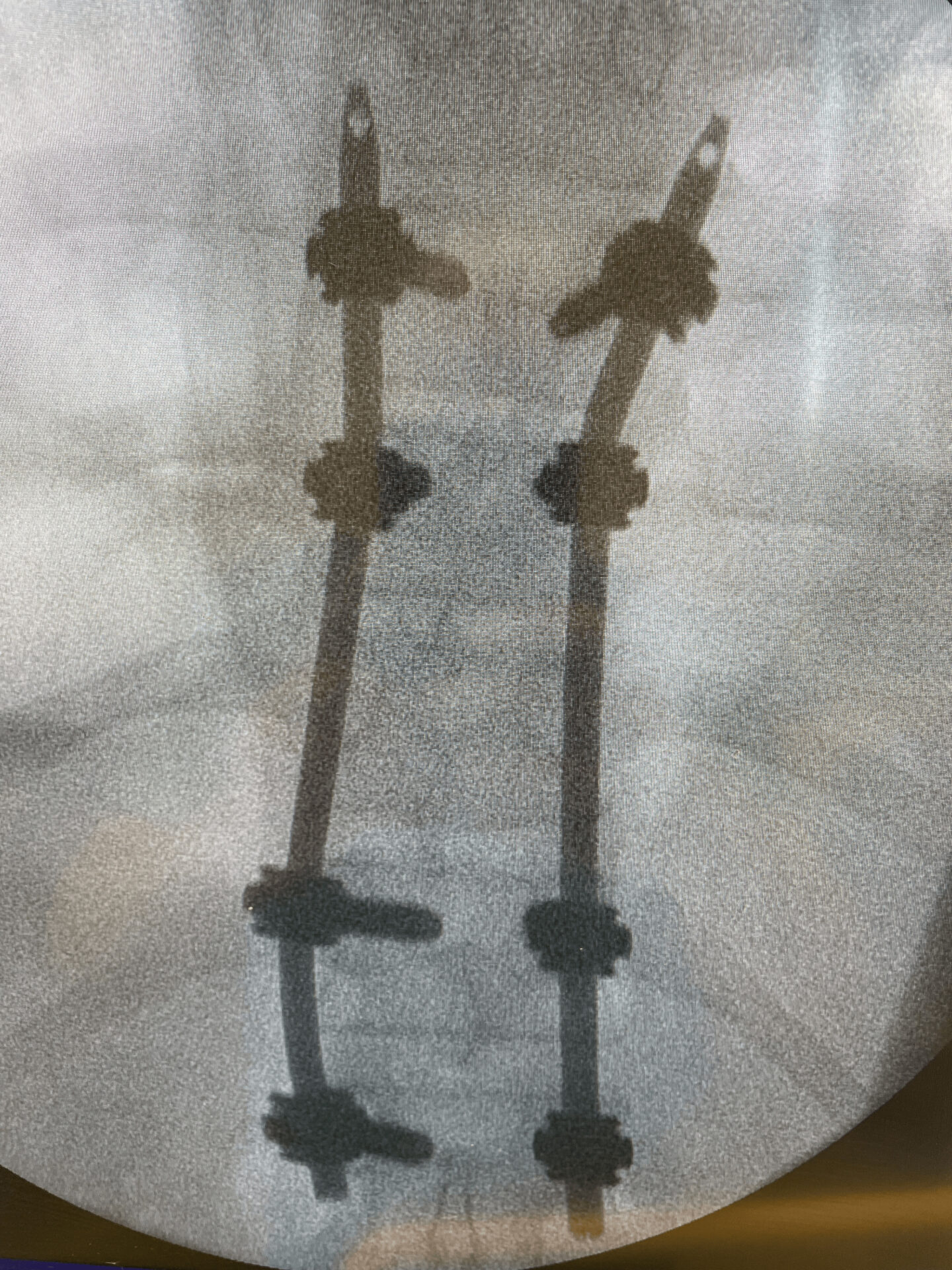
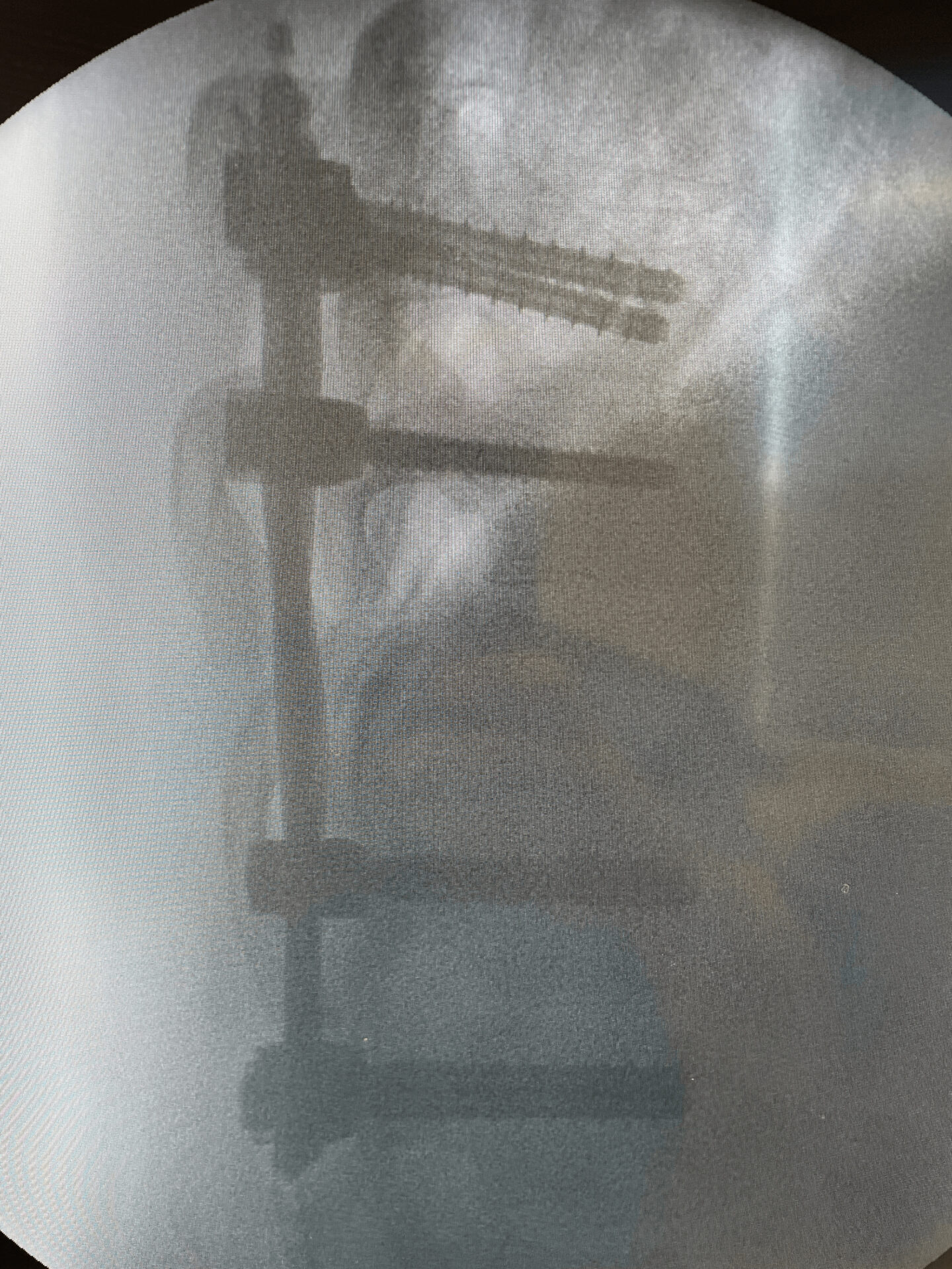
Radiographs after final fixation of the posterior construction and removal of the instrumentation.
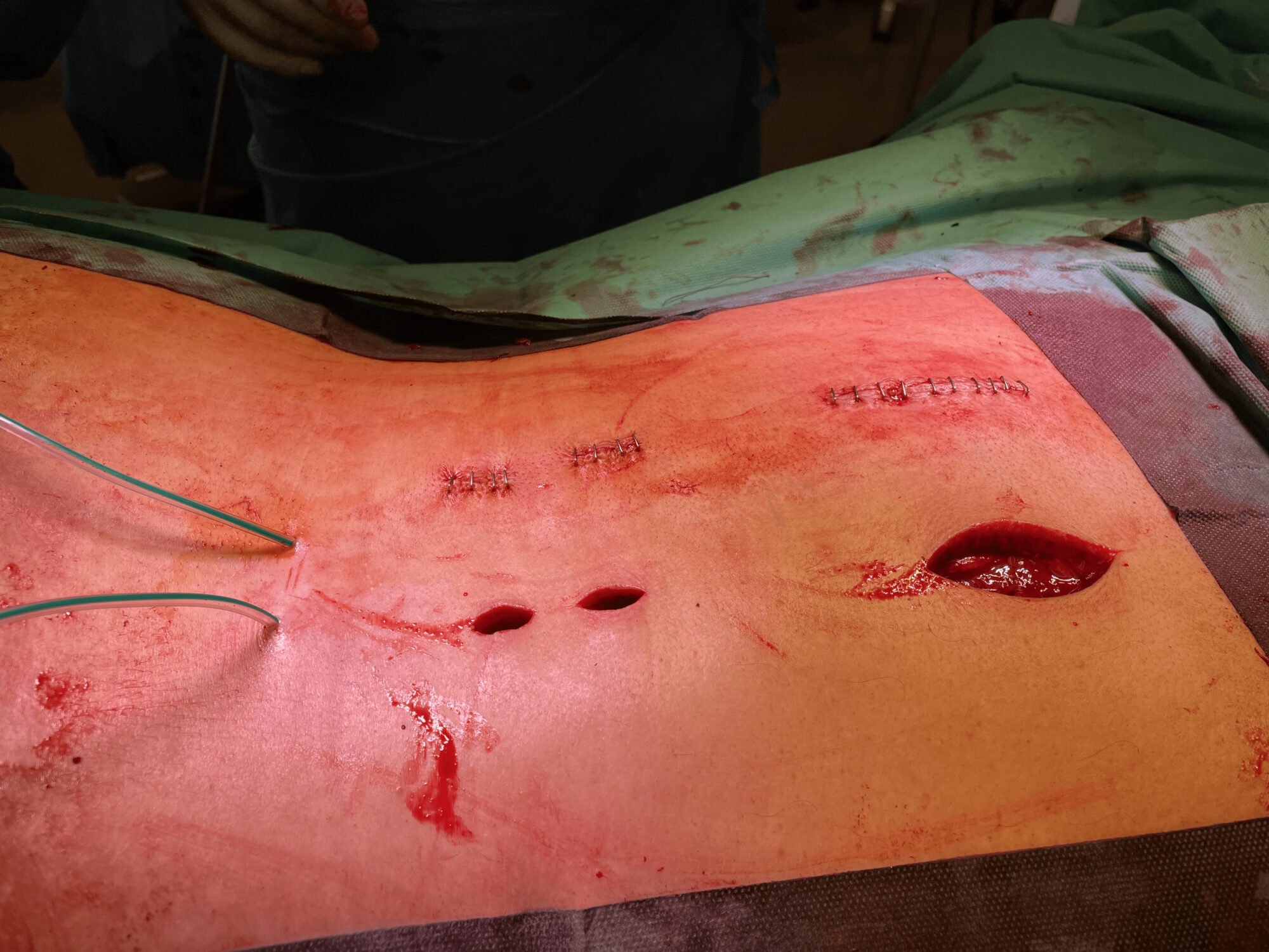
Closure of the surgical field

Post OP

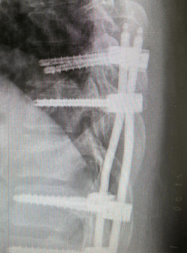
Post OP radiographs,
Sagittal and frontal views
- Intra OP Blood loss: 300 ml
- Duration of surgery: 90 min
- Later in the day of surgery a secondary bleeding occured resulting in a revision with 350 ml blood-loss.
- Patient was discharged from the hospital after 10 days


Published with the approval of Tino Beylich, MD


