E-Case report
DDD, Degenerative Disc Disease
Spinal Fusion L2 – S1Konstantinos Kafchitsas, MD PD

Pre OP

Clinical Case – Degenerative Disc Disease
Spinal Fusion L2 – S1
Neo ADVISE™
PD Dr. Konstantinos Kafchitsas
Asklepios Klinik Lindenlohe
Schwandorf
Germany


Patient Information:
83-year-old female patient with degenerative disc disease in lumbar region
Pain therapy at home resulted in no improvement
Diagnosed with:
- L3-4 spondylarthrosis
- L4-5 olisthesis with spinal canal stenosis, osteochondrosis
- L5- S1 lumboischialgia on the right side, and slowly develloping also on the left side
- Limping gait pattern
- Percussion pain deep lumbar over the spinous processes
- Positive facet compression test of the lumbar spine
- Lasègue’s sign positive on both sides from 30°
- No sensory motor deficit in any side
- Acute hemolytic anemia
- Postmenopausal osteoporosis
- Urinary tract infection
- Hip joint prosthesis left side
Planned Surgery:
- General anesthesia
- Open surgery, midline access
- PLIF expandable cages L2/3, L4/5, L5/S1
- Decompression L2/3, L4/5
- Posterior fixation L2 – S1
- Neo ADVISE™ 3D scanning


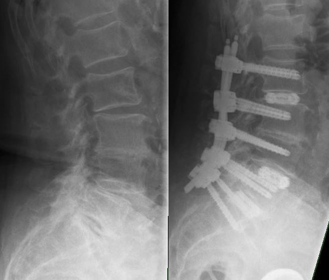
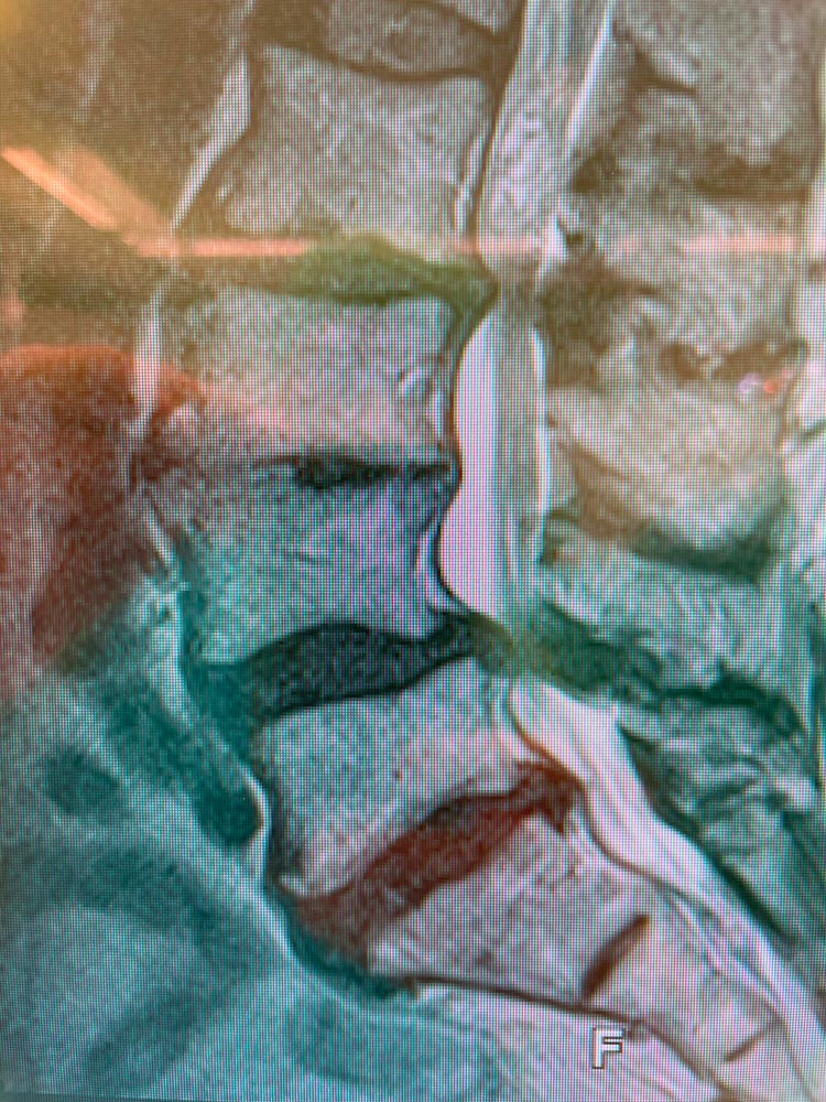
Pre OP standing radiographs & MRI, frontal and sagittal views.



Intra OP


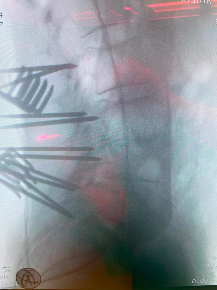
Midline access and short K-wires placement


Neo Pedicle markers to mark the intended screw trajectory. Decompression.
3 x PLIF expandable cages in L2/3, L4/5, K5/S1 (26x10mm, 3° Suzhou Kangli Orthopedics)
Bone graft – calcium phosphate cement (Neocement®)


Posterior fixation with Neo Pedicle Screw System™
Totally 10 pedicle screws were placed over the levels L2 – S1
-
L2 – 2 x Ø7.0x50 mm
-
L3 – 2 x Ø7.0x50 mm
-
L4 – 2 x Ø7.0x50 mm
-
L5 – 2 x Ø7.0x50 mm
-
S1– 2 x Ø7.0x45 mm


Visualizations of the shape of one of the rods, both the sagittal and the coronal planes, in the iPad screen as a result from using the Neo ADVISE™ 2 x titanium rods 130mm straight, cut to final 120mm length.


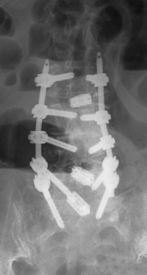
Radiographs after the final fixation of the posterior construction, and removal of the instrumentation
- Duration of surgery: 3h45min
- Post OP VAS: 4
- Patient was discharged from hospital after 13 days

Post OP
Radiographs after the final fixation of the posterior construction, and removal of the instrumentation

Post OP radiographs
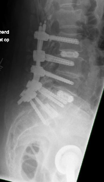
Radiographs, sagittal and frontal views at 3 months Post OP

Published with the approval of Dr. Konstantinos Kafchitsas


