E-Case report
Degenerative Disc Disease (DDD)
Spinal Canal Stenosis-Revision surgery
Posterior fixation T9 – L2David Schul, MD
Martin Reiser, MD


Pre OP

Clinical Case – Degenerative Disc Disease (DDD)
Spinal Canal Stenosis
Revision surgery
Posterior fixation T9 – L2
Neo ADVISE™
Dr. med. David Schul, Neurosurgeon
Dr. med. Martin Reiser, Neurosurgeon
InnKlinikum
Mühldorf, Germany


Dr. med. David Schul

Dr. med. Martin Reiser

Patient Information
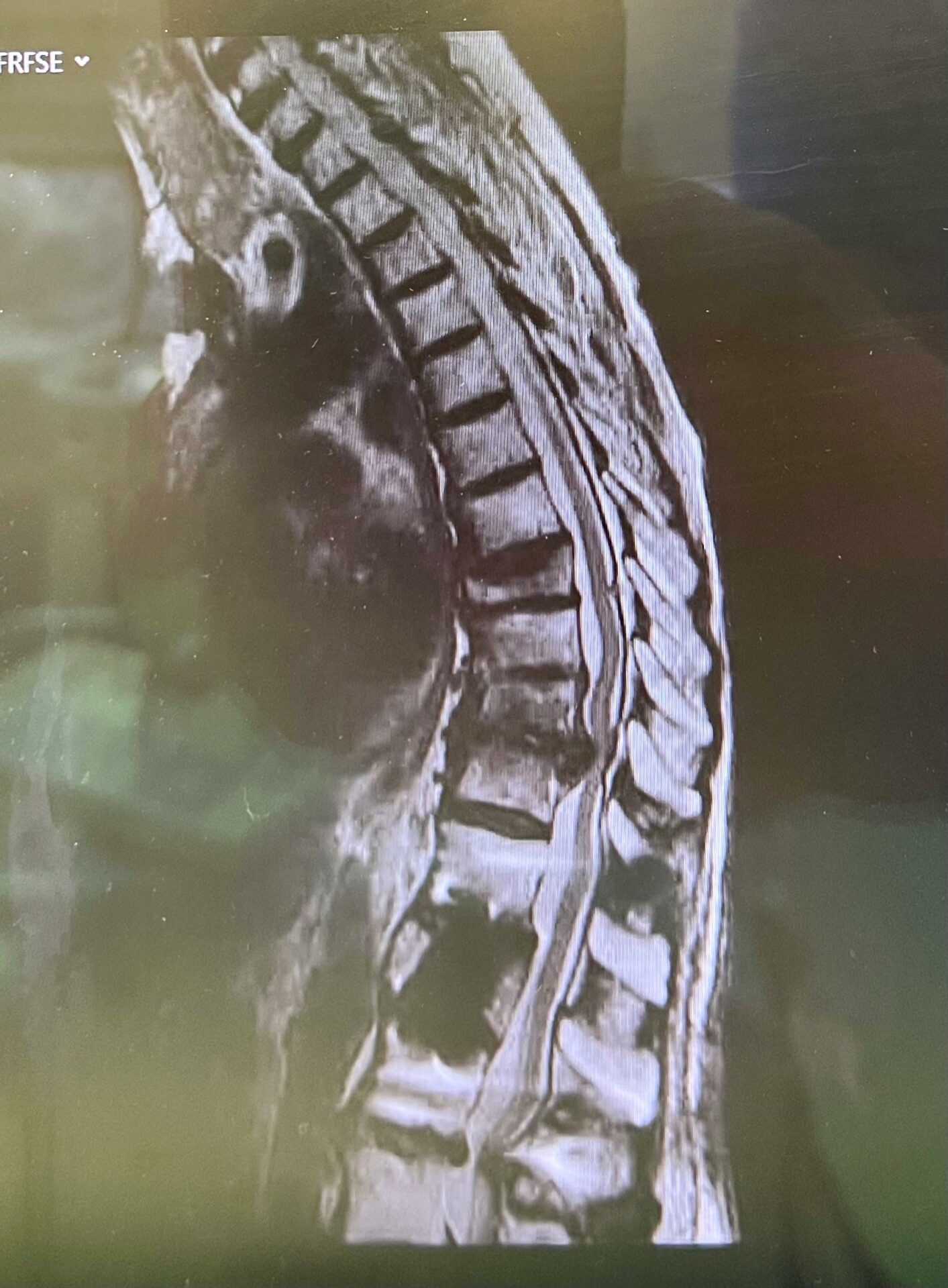
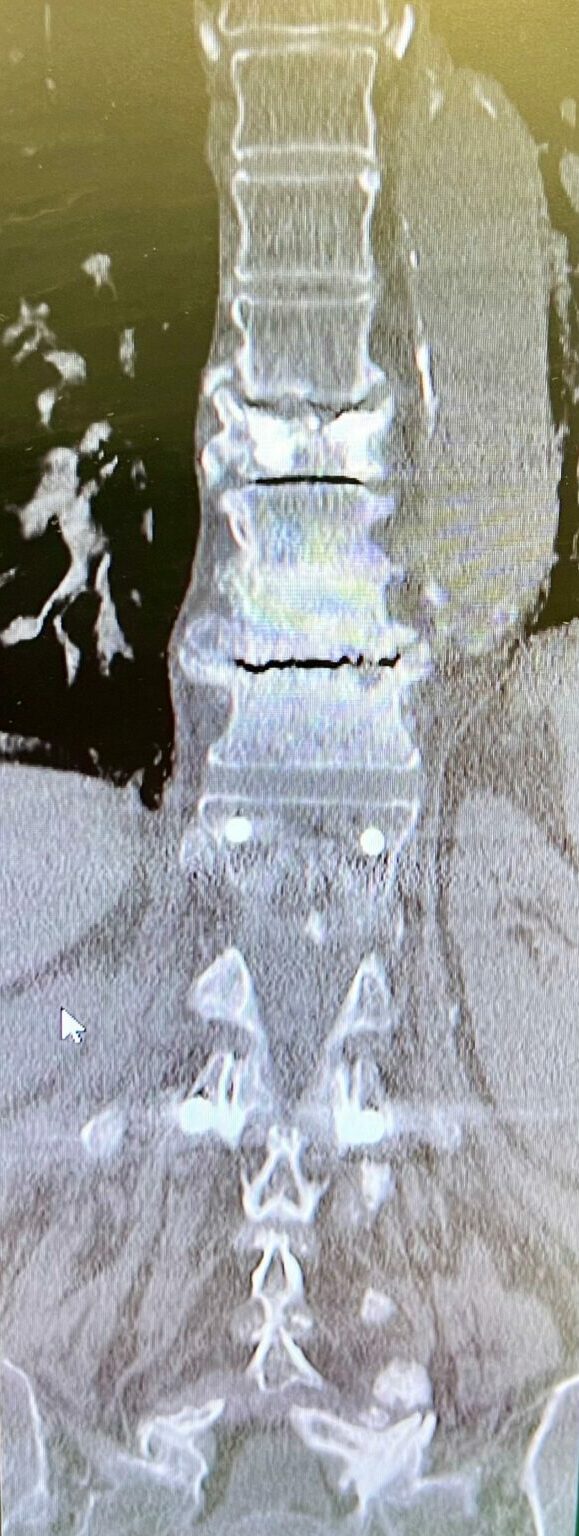
83-year-old female patient diagnosed with large calcified disc herniation and spinal canal stenosis in level T10/11.
The patient has earlier been treated with a L1 vertebral body replacement including dorsal stabilization T12 – L2. In addition, kyphoplasty in level T8.
Bone quality: Good
Pain (VAS): Minor pain (2-3)
Function (ODI): Moderate disability (35)
Neurological status: Paraparesis, patient couldn´t walk anymore
Hypertension
Planned Surgery:
-
- Revision surgery, removing of the dorsal instrumentation, pedicle screws in T12 and L2
- Open surgery, midline approach
- Posterior fixation T9 – L2
- Neo ADVISE™ to be used for a correct rod bending


Pre OP MRI & CTs, sagittal, frontal and axial views



Intra OP

Surgical procedure
-
-
- Patient placed in prone position
- General anesthesia – ITN & TIVA
- Open approach, midline cut
- Removal of old pedicle screws in level T12 & L2
-

Access and K-wires placement

- Costotransversectomy T9-11 with sequential removal of the sequestrum
- Laminectomy and bilateral decompression with microscope T10/11
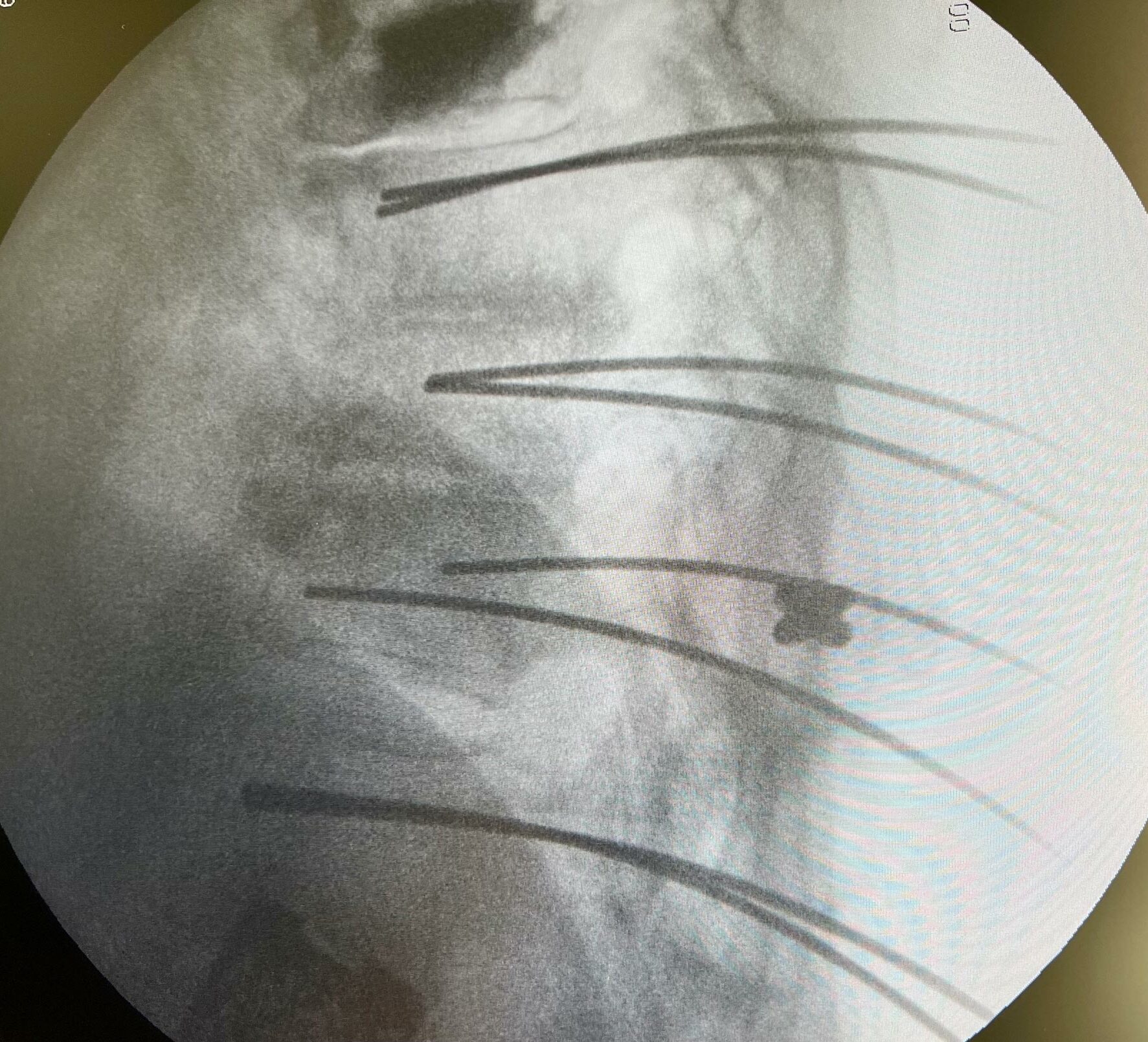
- K-Wire placement (lateral) in levels T9-T12 and L2 using navigation BrainLab
-
Posterior fixation with Neo Pedicle Screw System™
Totally 10 pedicle screws placed over the 5 levels T9 – L2
- T9 2 x Ø6.0x40mm
- T10 2 x Ø6.0x40mm
- T11 2 x Ø6.0x45mm
- T12 2 x Ø6.0x50mm
- L2 2 x Ø7.0x50mm


Usage of Neo ADVISE™

ADVISE™ scene analysis


Results from using the Neo ADVISE™ in both sides, with visualizations of the rods in both the sagittal and the coronal planes shown in the iPad screen.
Rod selection:
2x Titanium rods 160mm straight
ADVISE™ 152/155mm


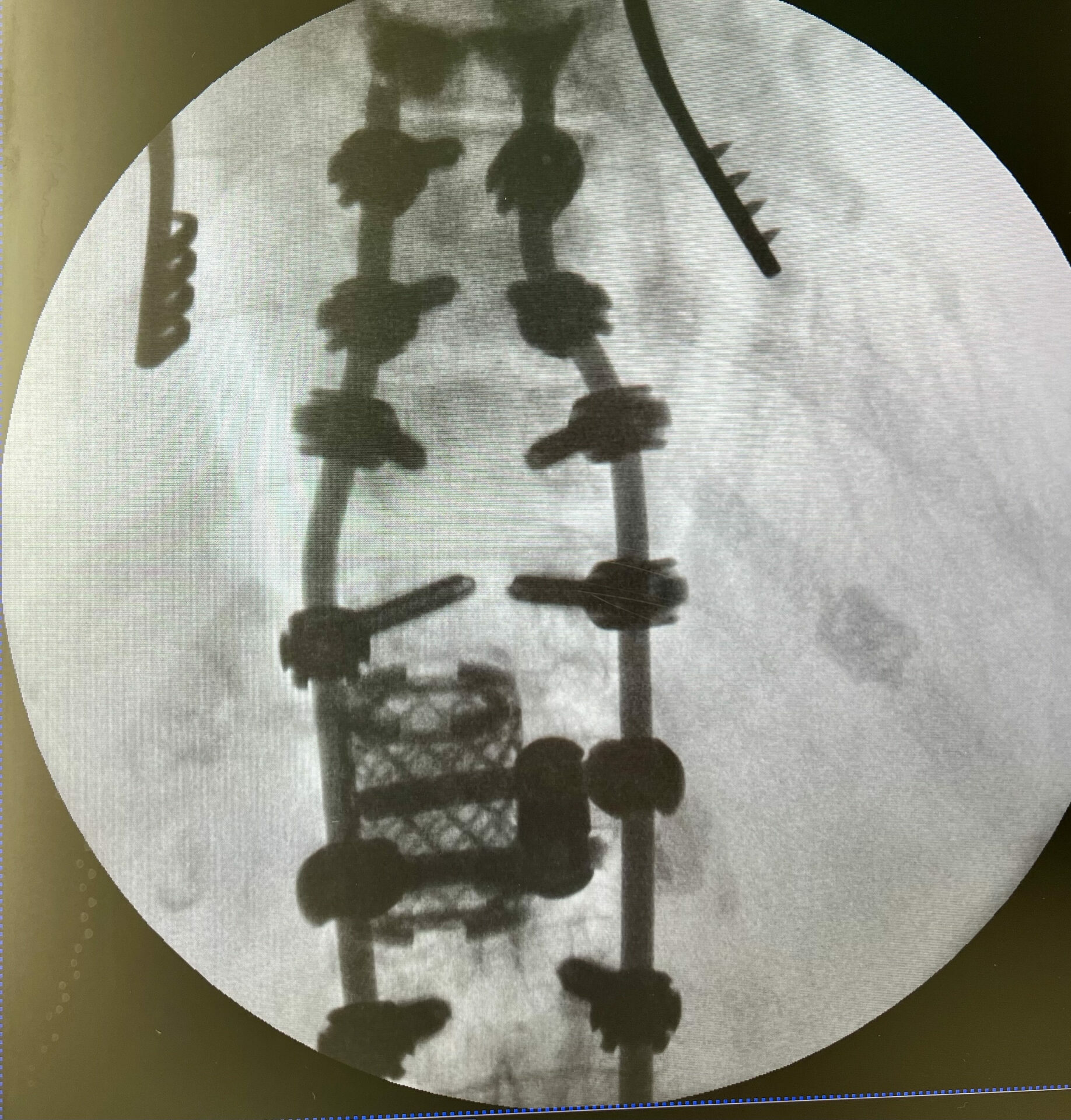
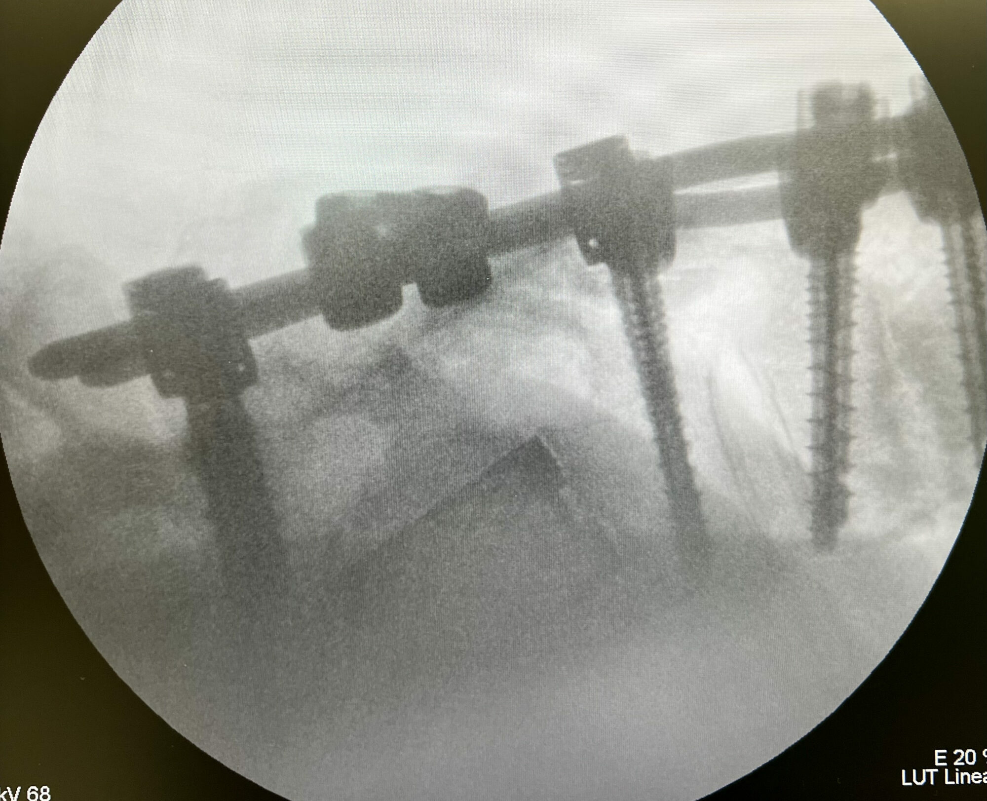
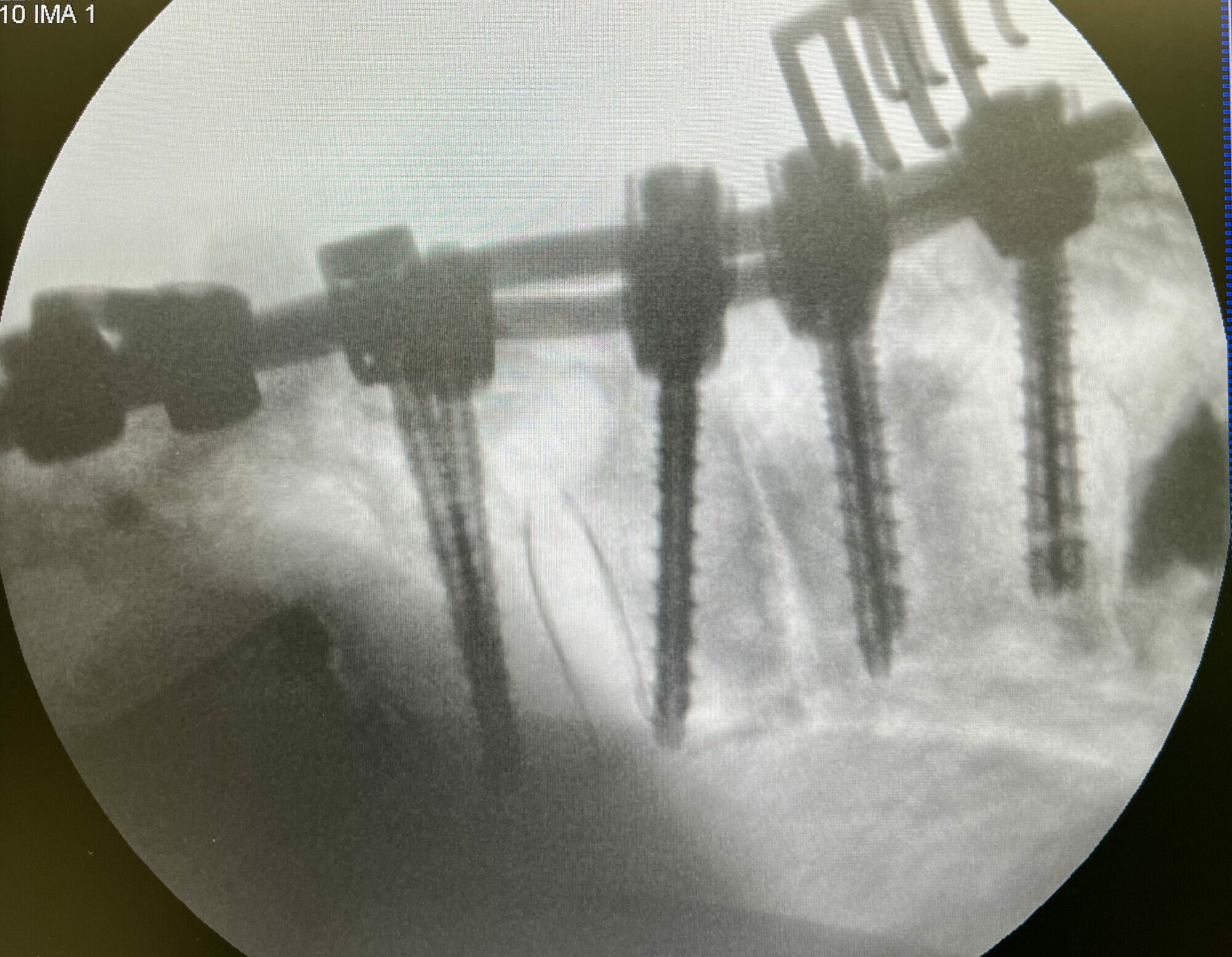
After the final fixation of the posterior construction and removal of the instrumentation.
A cross connector was inserted to increase stability.
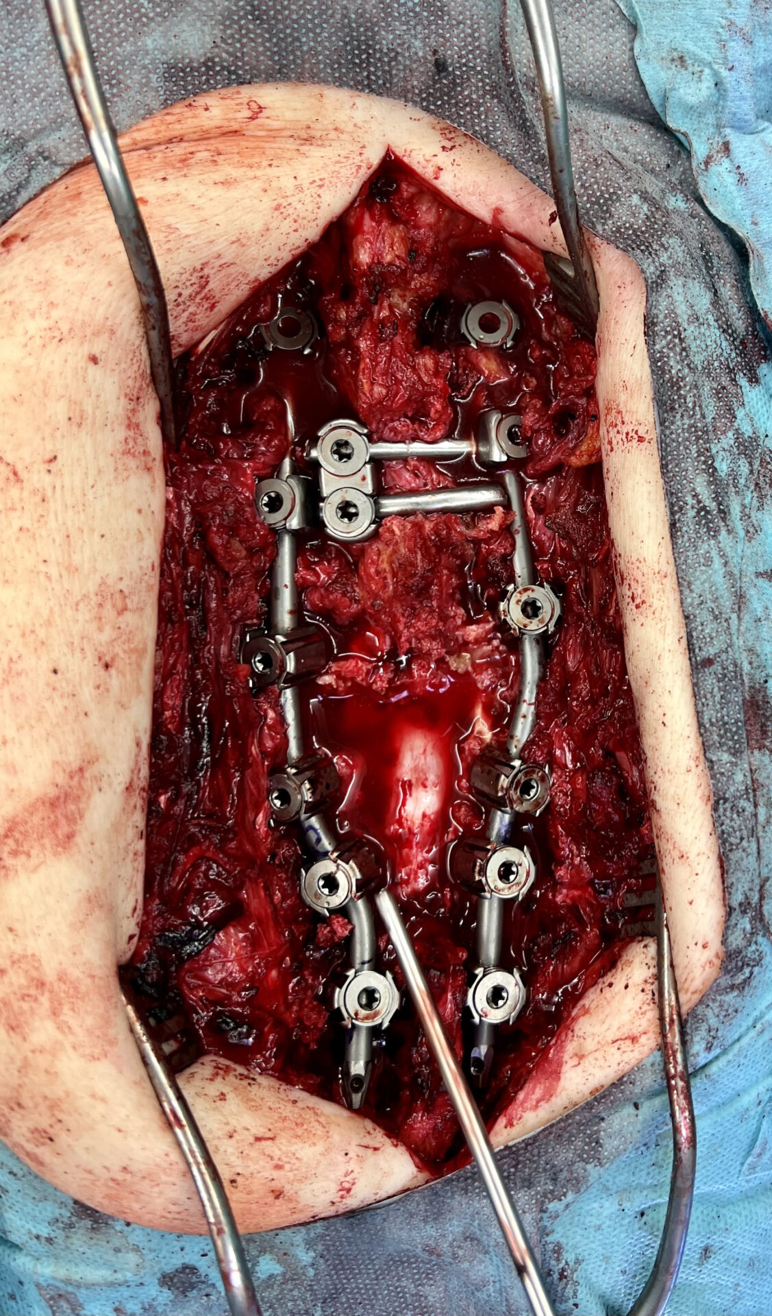

Radiographs after final fixation of the posterior construction and removal of the instrumentation.

Post OP

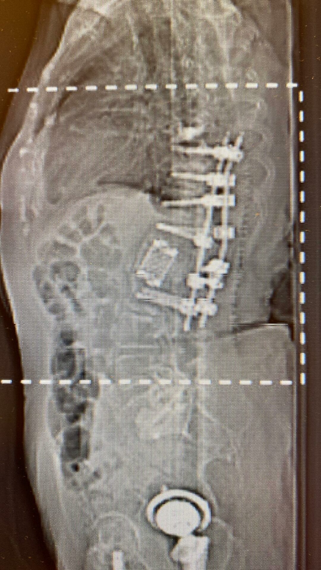
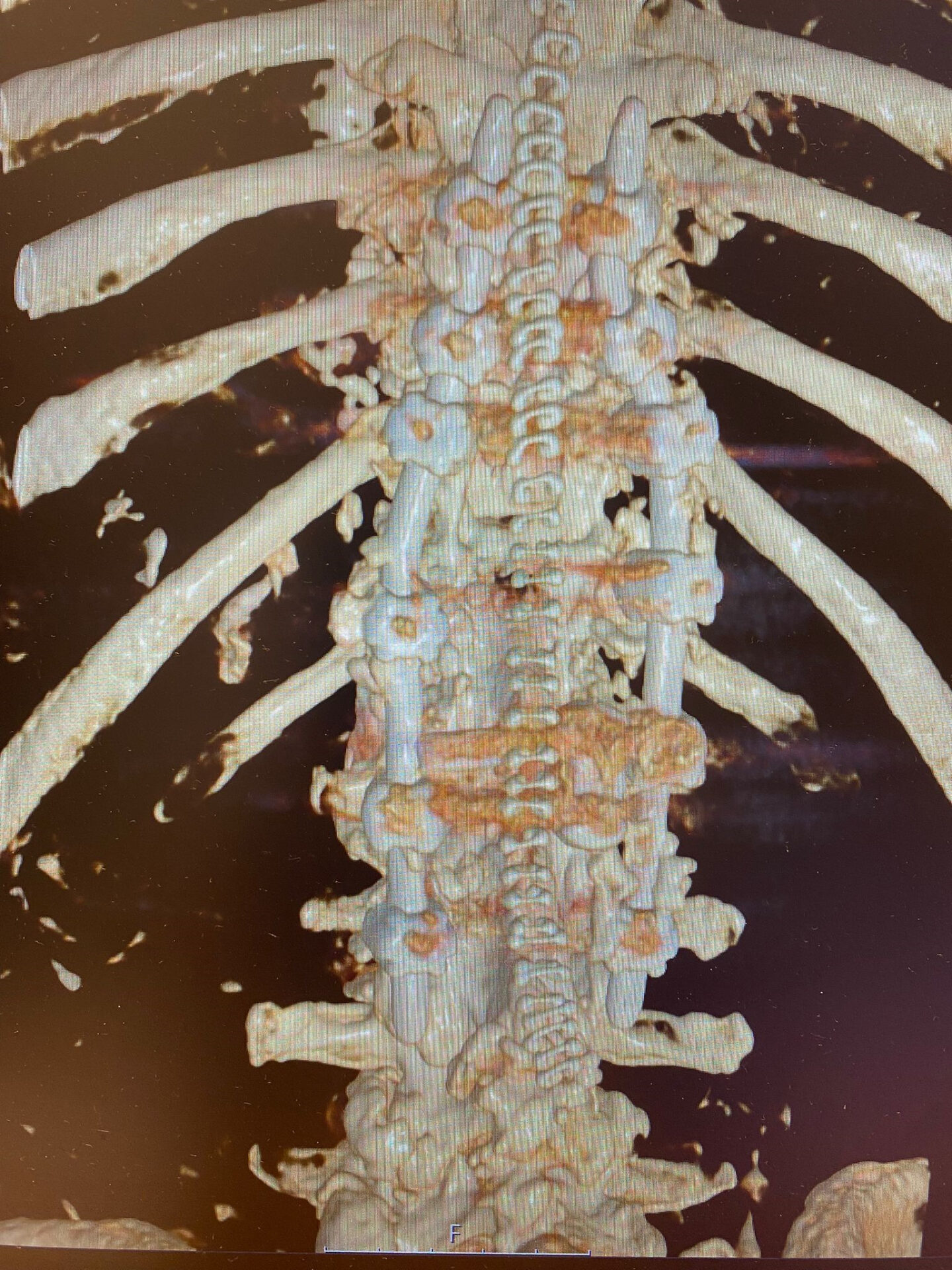
Post OP CT scans
- Intra OP Blood loss: ca. 500 ml
- Duration of surgery: 500 min
- Pain (VAS): 2-3
- Significantly better neurological status, the patient could leave the hospital walking
- Patient was discharged from the hospital after 7 days
-

Published with the approval of Dr. David Schul and Dr. Martin Reiser


