E-Case report
Multiple Myeloma T10-L2 Dr. med. Juan José Correa Gámiz
Dr. med. Francisco López Caba
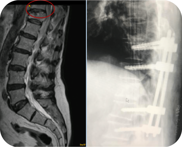
Pre OP

Clinical Case – Multiple Myeloma
Dr. med. Juan José Correa Gámiz
Dr. med. Francisco López Caba
Hospital Virgen de La Luz, Cuenca
Cuenca CUENCA
Spain


Patient Information:
- 75-year-old male patient
- History of hypertension, diabetes mellitus, prostate cancer in remission
- Patient attended trauma consultations for omalgia, shoulder pain left side since 4 months, and lumbar pain, both with mechanical characteristics
- Patient reported fever, but no weight loss
- Percutaneous biopsy of the T12 vertebra diagnosed plasmacytoma
- Diagnosis by Multiple Myeloma after complementary hematological laboratory tests and PET-CT (uptake in proximal femurs, ischiopubic branches, left clavicle, left 3-4º rib arch).

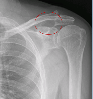
Pre OP radiographs
- Radiograph shows a lytic lesion on the left scapula

Radiograph shows a lytic lesion on the left scapula

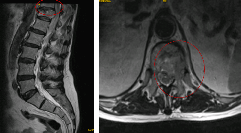
- At the thoraco-lumbar level, a slightly wedging of the upper endplate of T12 can be seen
- Scapular and lumbar MRI: showing lytic lesions in the left scapula and left side of T12 vertebra with involvement of the posterior wall and pedicle with spinal compression that does not generate clinical symptoms

Pre OP situation & Surgical planning:
- Mechanical lumbar pain without associated neurological symptom
- Instability classification scale (SINS): 13
- Decision to perform percutaneous fixation without arthrodesis given the absence of spinal compression and with the benefit of starting early radiotherapy treatment
- No alteration of the sagittal balance
Intra OP

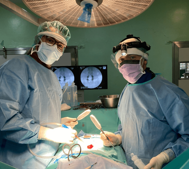
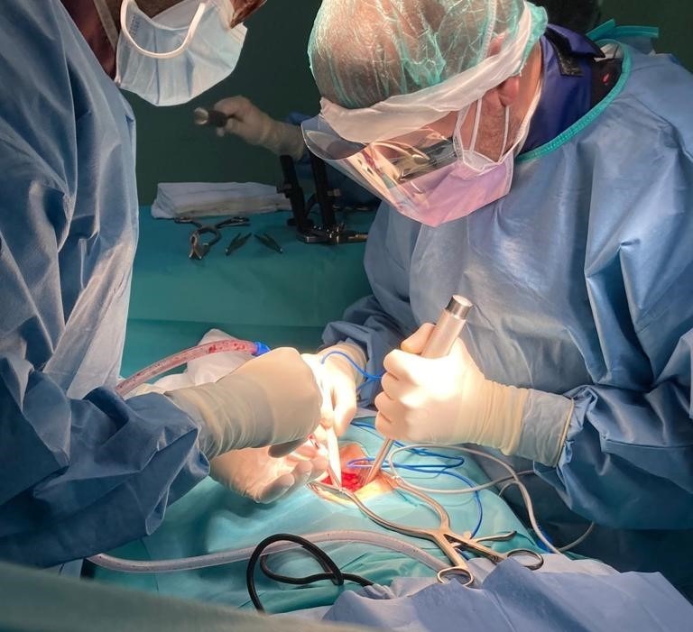
Surgical procedure
- General anesthesia
- Percutaneous approach
- Fixation T10-L2
- Neo Pedicle Screw System™
- 8 x Ø6.0 x 45mm pedicle screws
- Skin closure of incisions with staples
- Time of surgery: 1h40

Post OP
Post OP outcomes
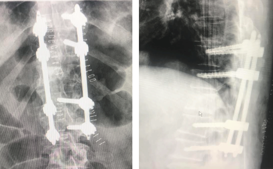
- No lumbar pain 2 days after surgery
- Ambulation without assistance, and with no pain
- No neurological symptoms
- Discharged from the hospital ward 48h after re-evaluation by the department of hematology
- Started chemotherapy and radiotherapy 7 days after surgery
- No surgical wound complications

Follow Up at 15 months
- At this time, 76 years old, with progress of the multiple myeloma despite the treatment with radio- and chemotherapy
- No back pain of mechanical characteristics
- No failure of the osteosynthesis material (pedicle screws or rods)
- The patient is followed biannually in an outpatient trauma clinic
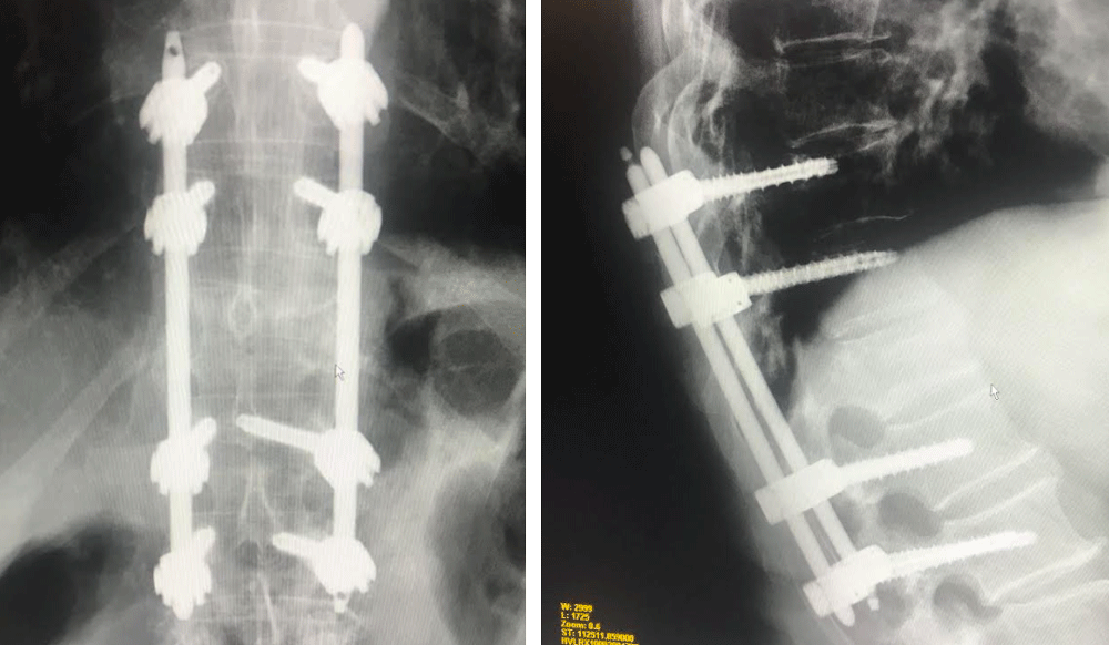
Radiographs at 15 months postoperatively, coronal and sagittal views

Published with the approval of Dr. med. Juan José Correa Gámiz & Dr. med. Francisco López Caba


