E-Case report
High-energy Trauma Fractures T8 & T11
Posterior fixation T4 – L1Eckard Hölscher, MD

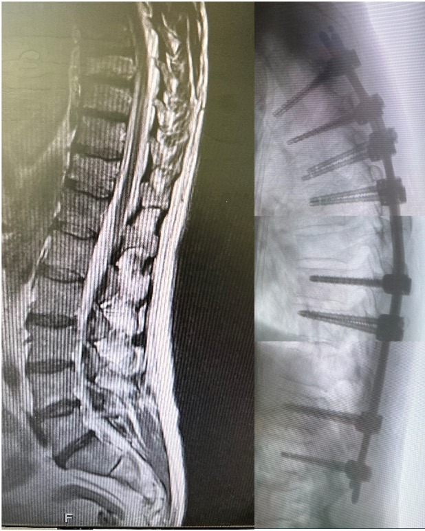
Pre OP

Clinical Case – High-energy Trauma Fractures T8 & T11
Posterior fixation T4 – L1
Neo ADVISE™
Eckard Hölscher, MD
Orthopaedic/Trauma Surgeon
Helios Klinikum Berlin Buch
Berlin
Germany


Patient information
Male 56 years old patient, construction worker who was admitted to the emergency department after a fall at work.
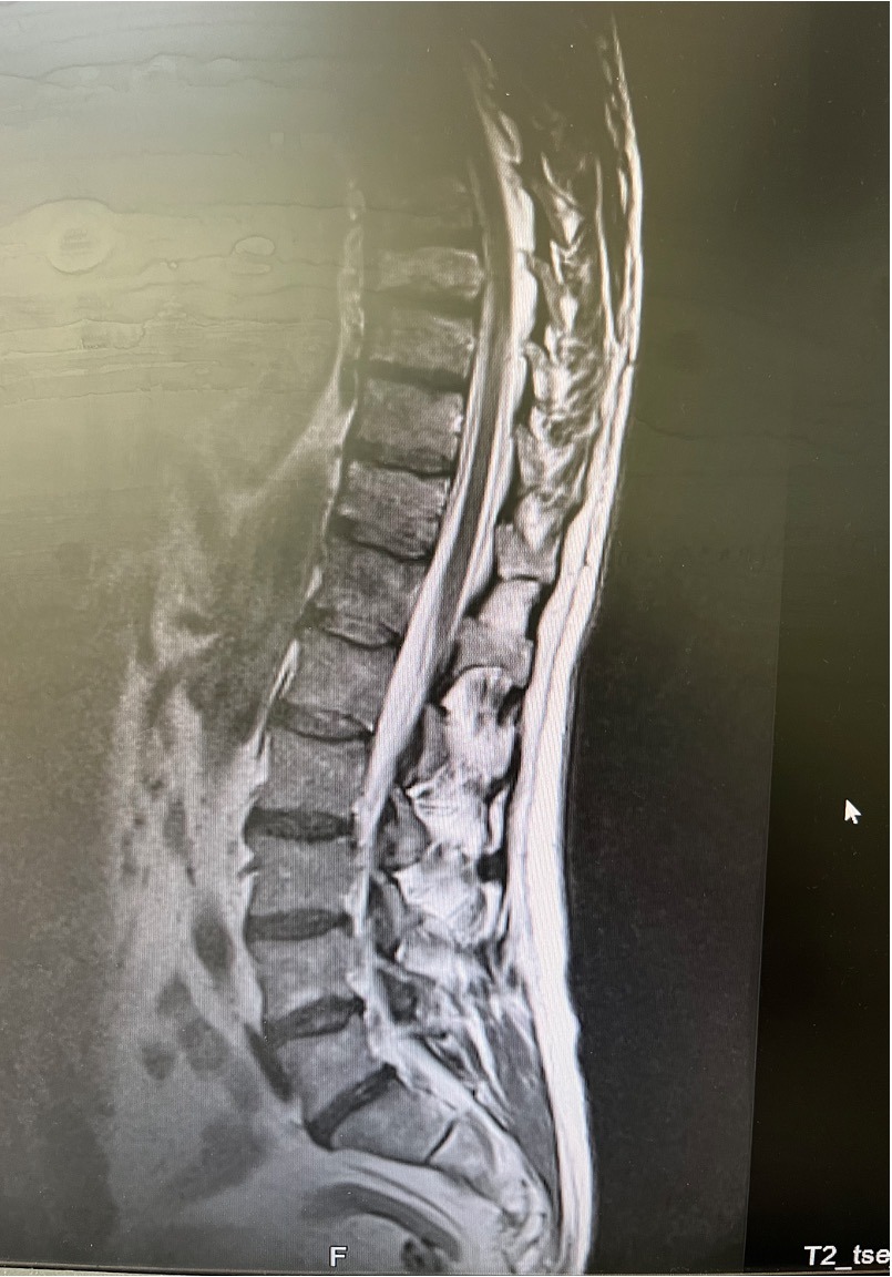
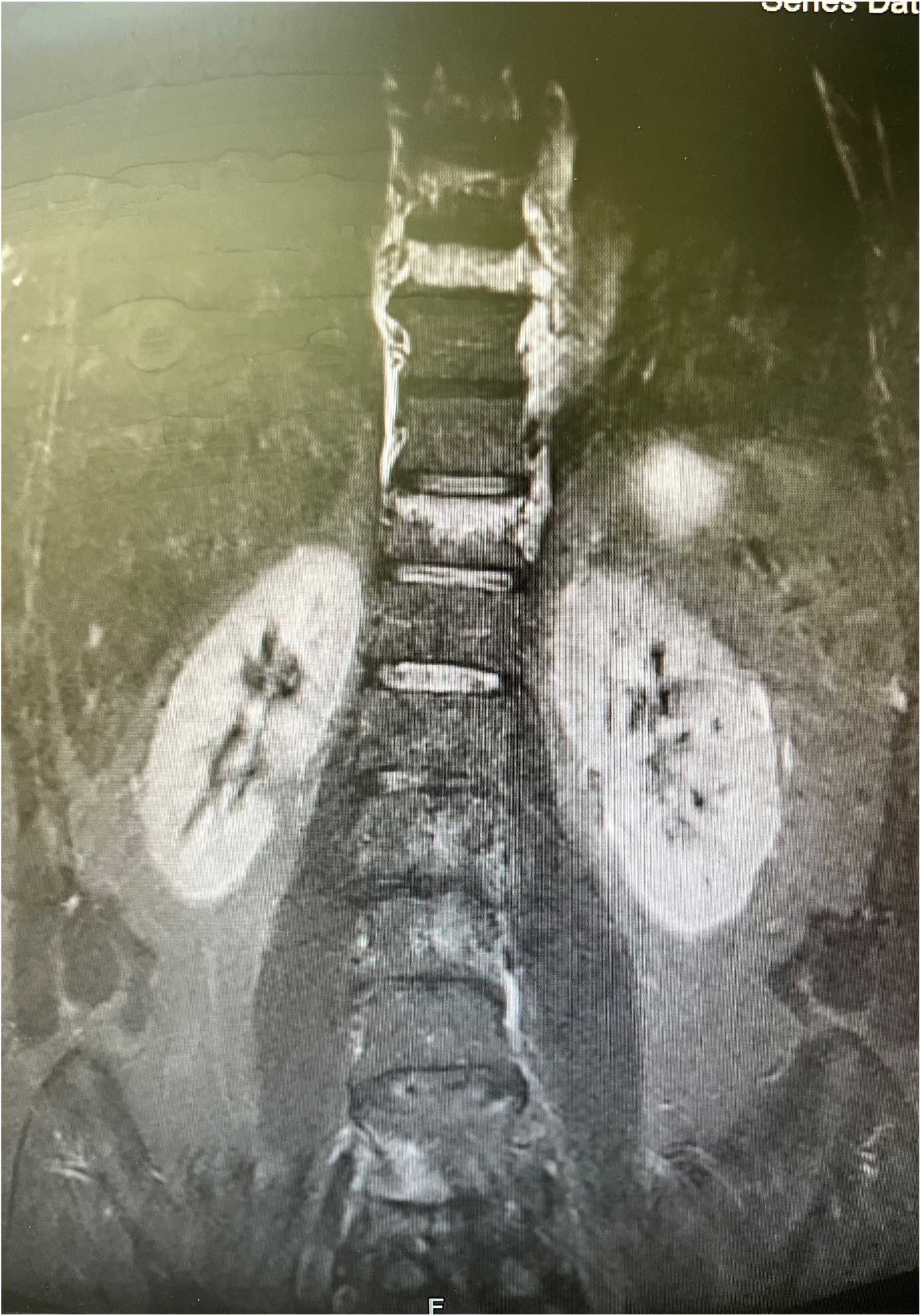
Polytrauma, multiple fractures: Vertebral fractures in
T8 – Endplate impression fracture
T11 – Burst fracture, type B
Fibula fracture
Calcaneus fracture
Neurological status: OK
Good bone quality
Planned Surgery:
- Posterior fixation, long construction (10 levels) T4 – L1
- Percutaneous MIS surgery
- Neo ADVISE™ , marker option, to be used


Pre OP MRIs
Sagittal & frontal views

Intra OP

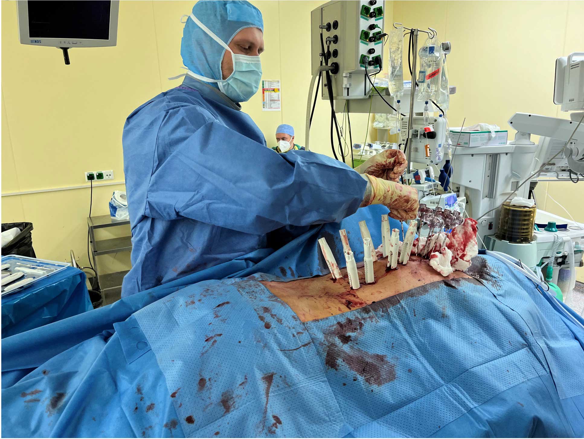
Pedicle screws are placed guided by the K-wires
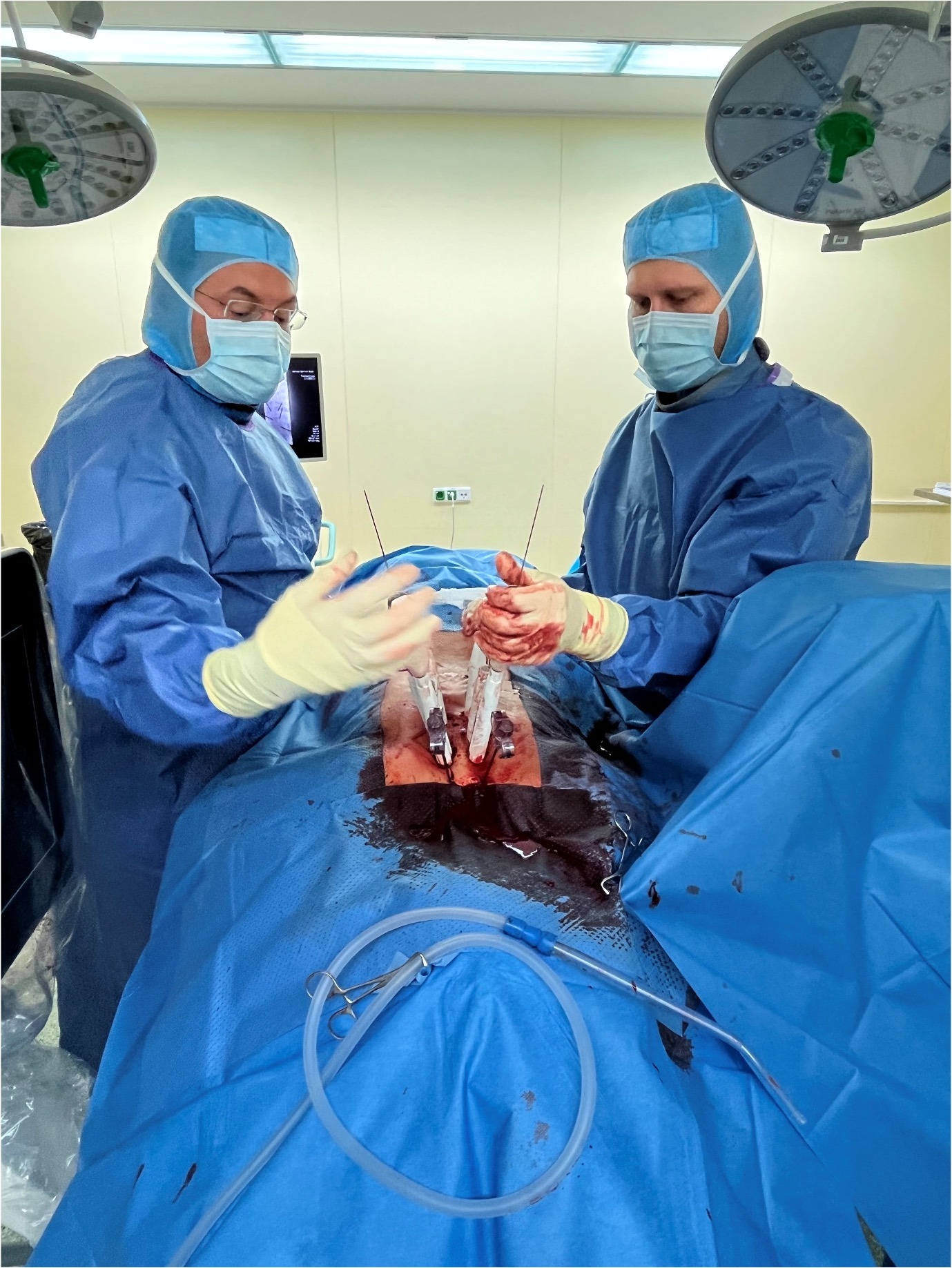


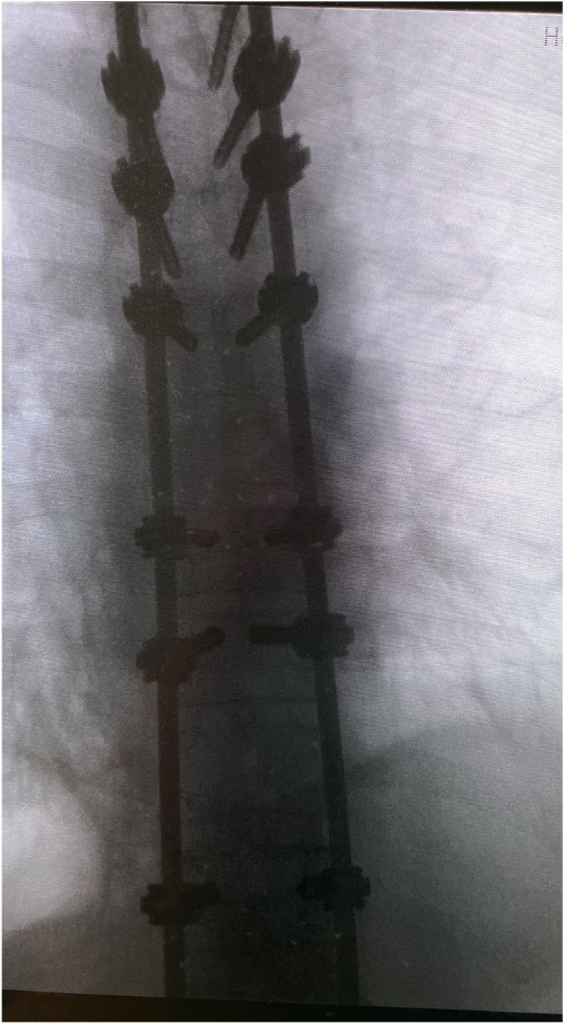
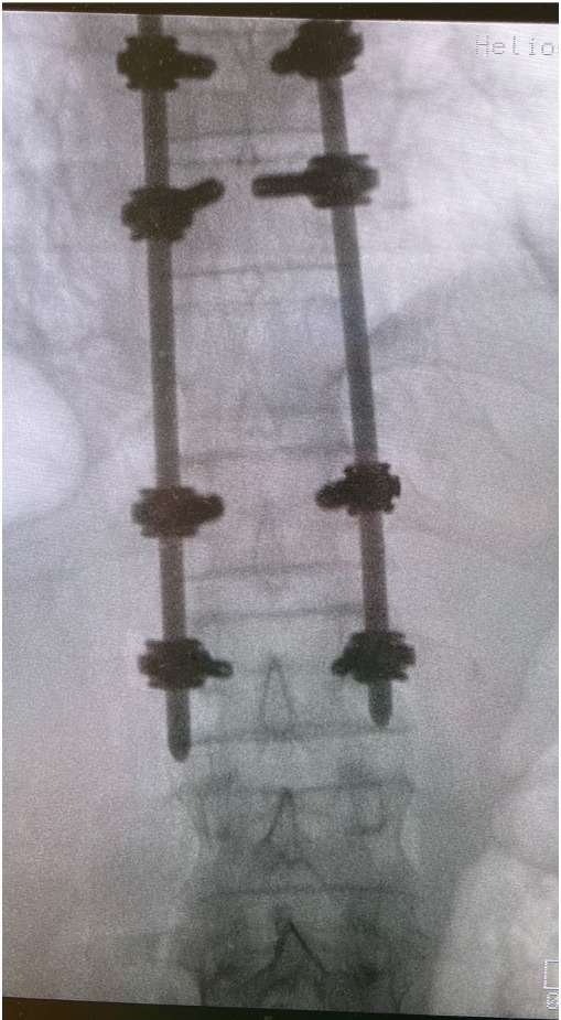
Posterior fixation, long construct, with the Neo Pedicle Screw System™
Totally 16 pedicle screws placed over the levels T4 – L1 (no screws in the fractured levels T8 and T11)
- T4 – 2 x Ø4.5×45 mm
- T5 – 2 x Ø4.5×45 mm
- T6 – 2 x Ø5.0×45 mm
- T7 – 2 x Ø5.0×40 mm
- T9 – 2 x Ø6.0×50 mm
- T10 – 2 x Ø6.0×55 mm
- T12 – 2 x Ø7.0×55 mm
- L1 – 2 x Ø5.0×50 mm
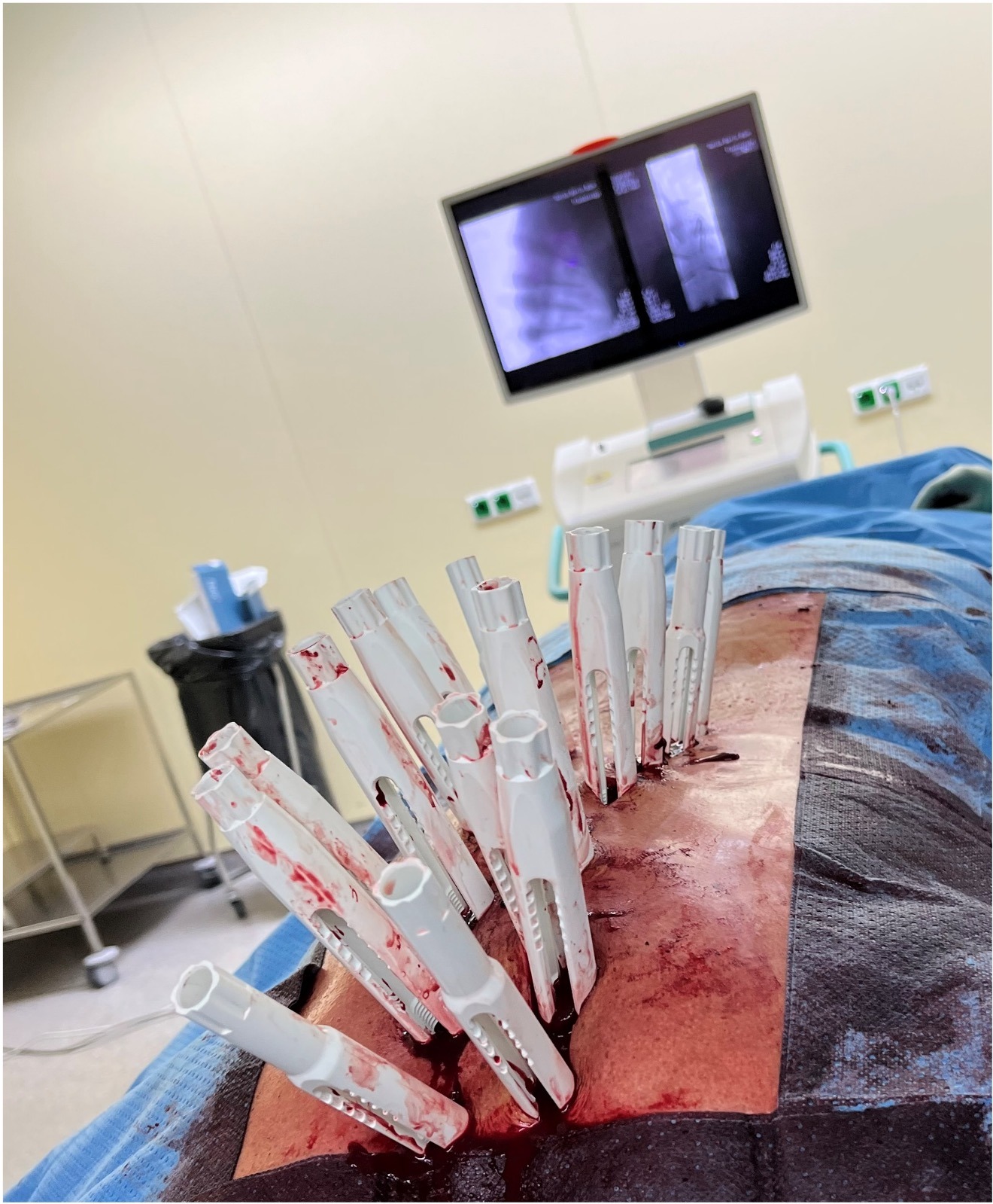
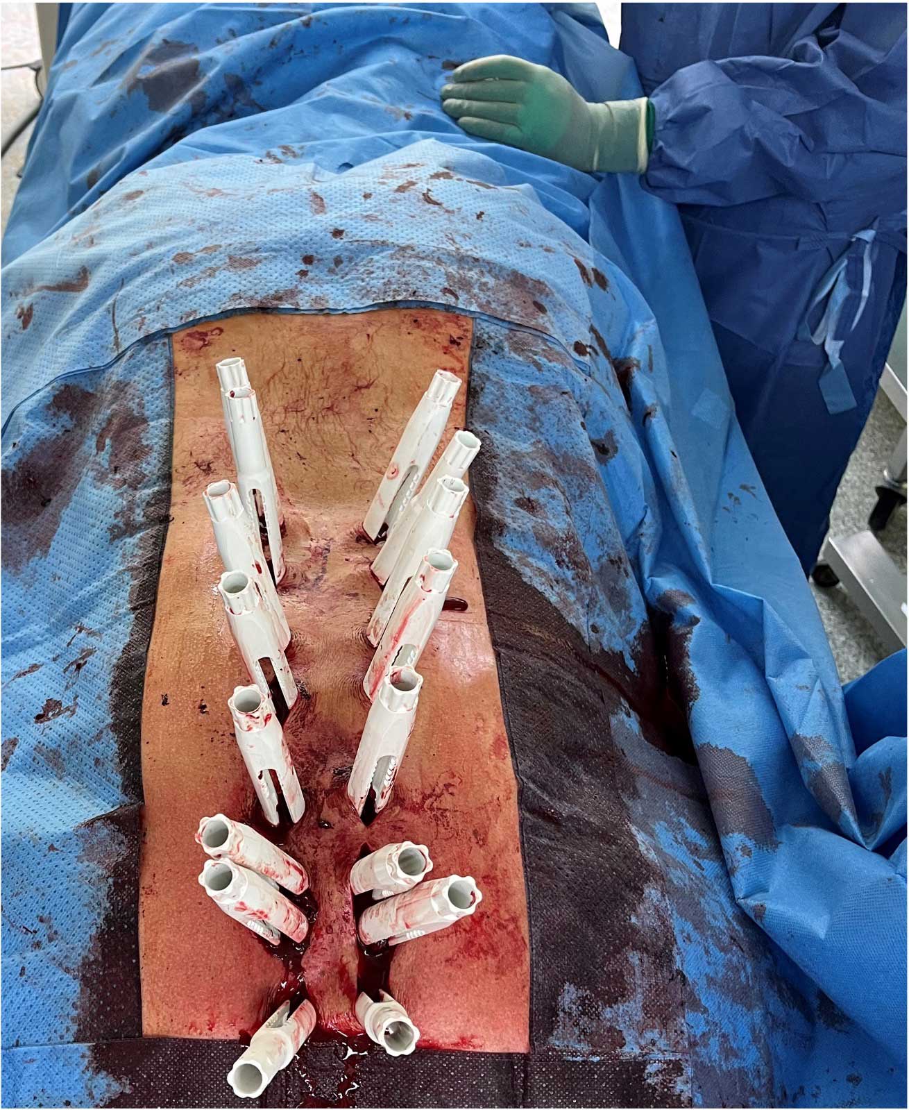


Neo ADVISE™
Marker Detection Method Markers are attached onto the screw guides.
The Marker Detection method works for scanning the guides on both sides of the spine at the same time.
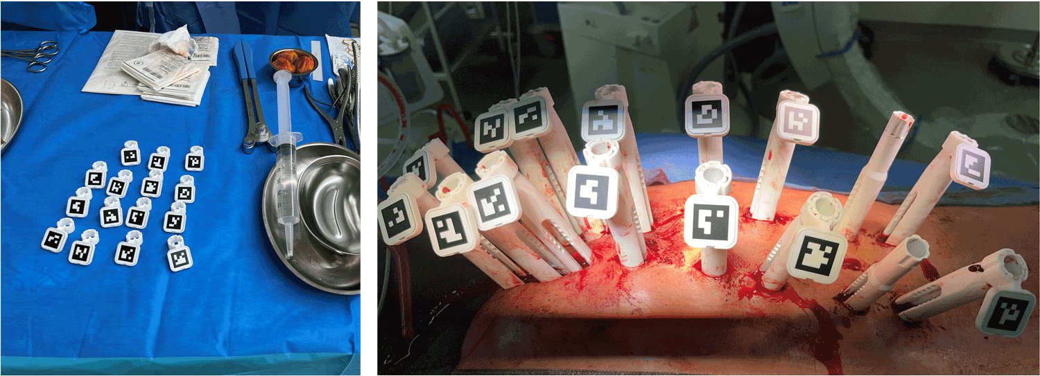
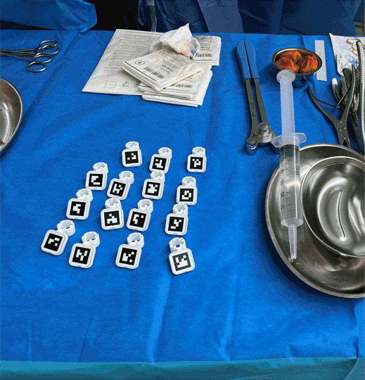
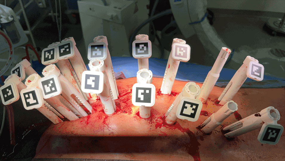


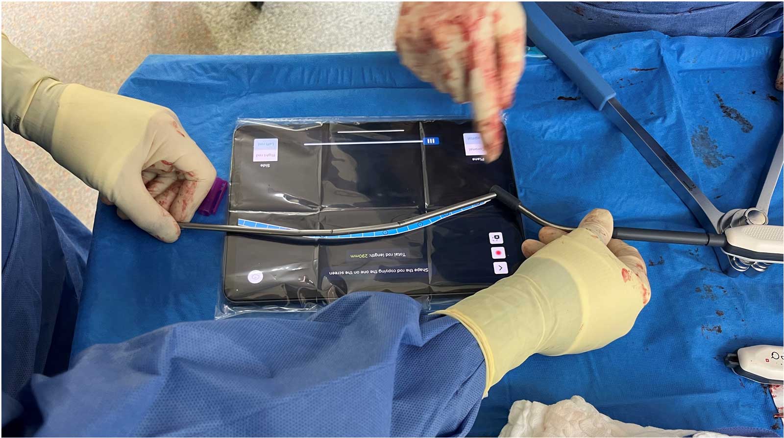
Mapping the markers, using the iPad screen
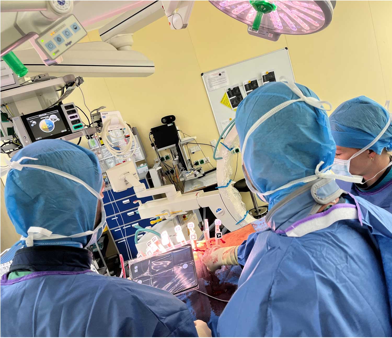
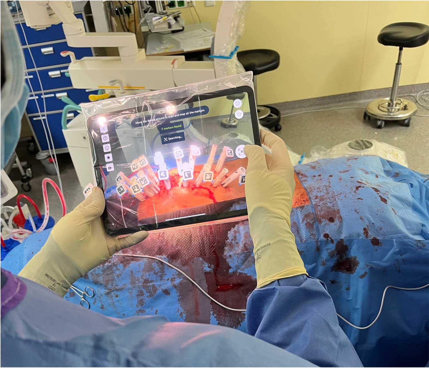


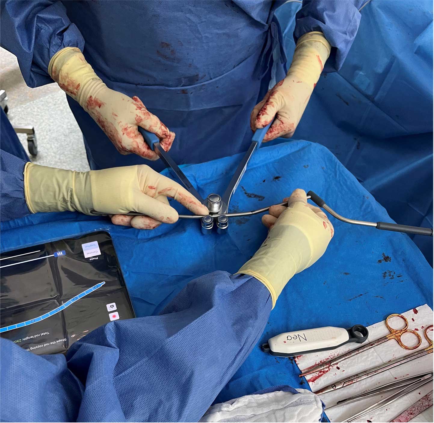
Bending of the left side rod, 300mm straight, by using the visualization of the rod in the sagittal plane shown in the iPad screen.


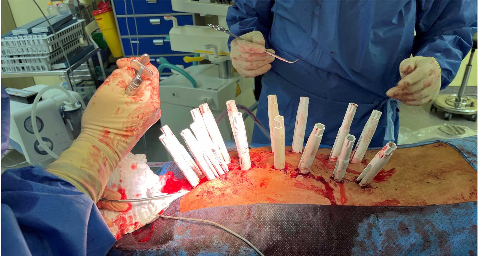
Placing left side rod

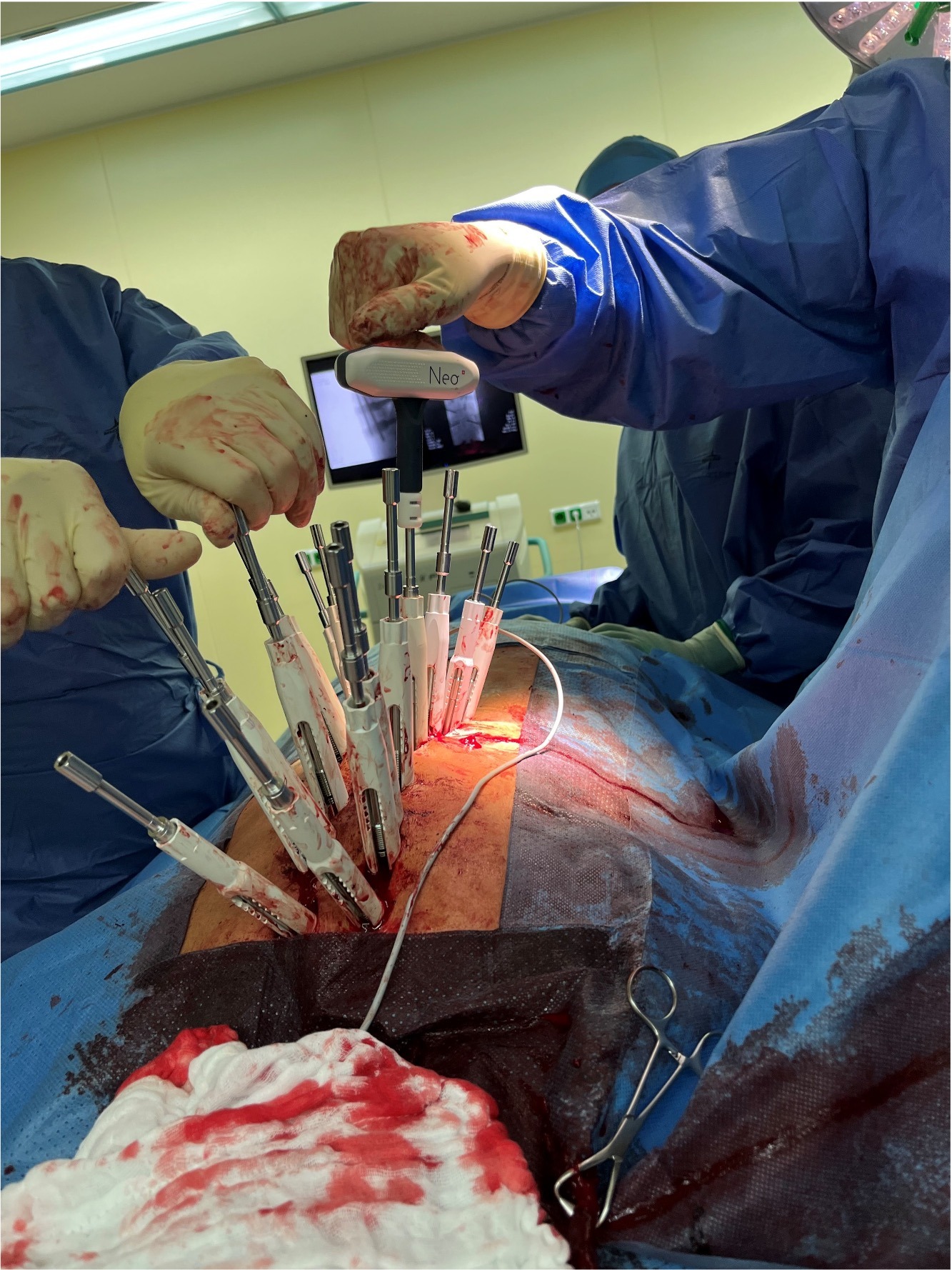
Pre-fixation of the set screws, using the torque limiter attached to the handle.


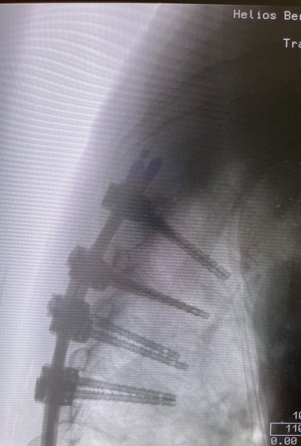
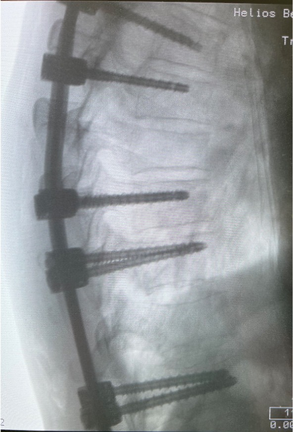
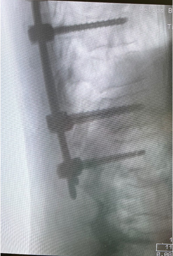
Radiographs, sagittal view, after final fixation of the posterior construction and removal of the instrumentation.

Post OP

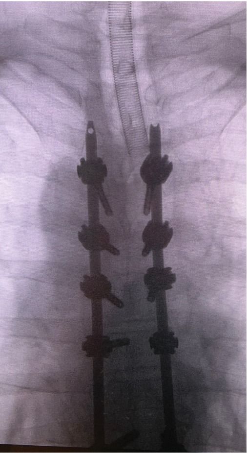
Post OP radiographs, frontal
- Intra OP Blood loss: 700 ml
- Duration of surgery: 180 min
- Patient was discharged from the hospital after 13 days

Published with the approval of
Eckard Hölscher, MD


