E-Case report
Minimally Invasive AIS SurgeryDr. Alejandro Peiró García
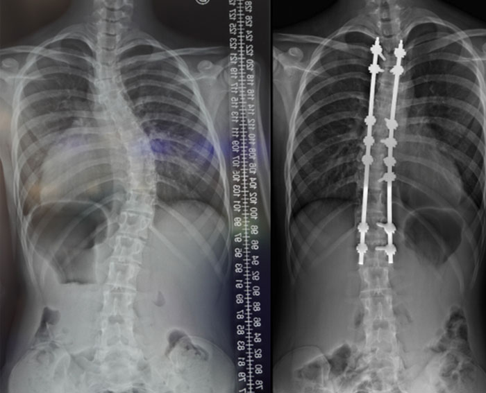
Pre OP

Clinical Case – Minimally Invasive AIS Surgery
Alejandro Peiró García , MD
Orthopaedic and Trauma surgeon
University Hospital Sant Joan de Déu
Barcelona, Spain


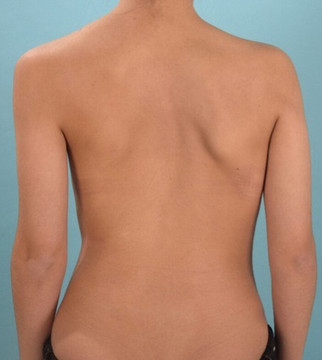
Patient Information
Adolescent Idiopathic Scoliosis (AIS) in a 16 years old female patient
- Flexible AIS with main thoracic right curvature
- Single curve with apex at T9
The challenge of the surgery will be to:
-
fuse a minimum number of vertebrae trying to avoid motion restriction
-
reduce the soft tissue damage in a traditionally aggressive surgery


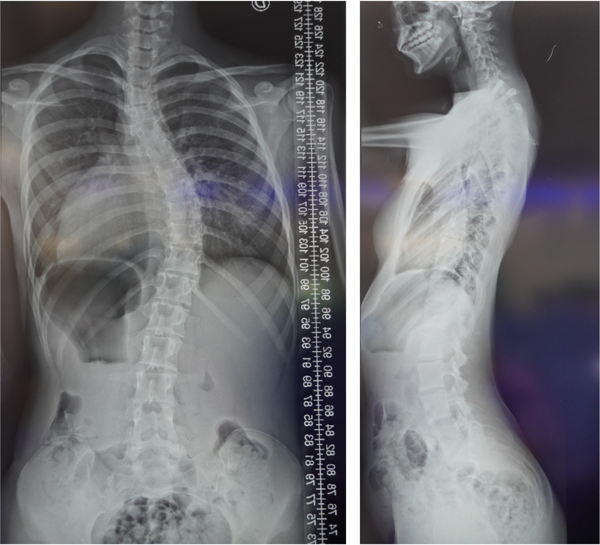
Pre OP
EOS standing radiographs
frontal & sagittal views


Surgical Planning
Type: Lenke 2B (Modifier B)
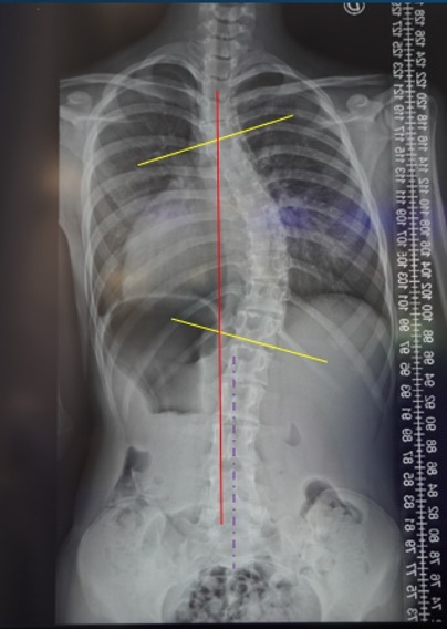
| Pre op | Correction Objective | |
|---|---|---|
| Cobb angle | 50° | <10° |
| Thoracic Kyphosis (TK) | -5° | >10° |
| Lumbar Lordosis (LL) | 47.3° | same |


Surgical Planning
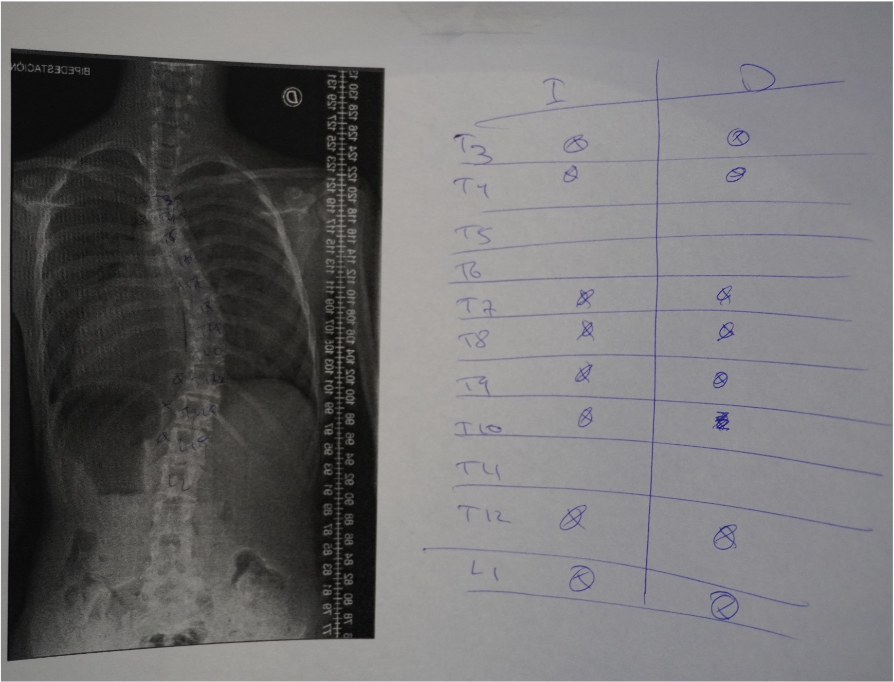
- Skin incision
- High thoracic (T3 -T4) percutaneously
- Apex (T7 – T10) midline open
- Bottom (T12 – L1) midline open
- Access & facetectomies
- CT screw navigation – Philips ClarifEye™
- K-wires & pedicle markers positioning
- T3 – L1 screw insertion – Neo Pedicle Screw System™
- Neuromonitoring check
- Rods insertion
- Postero-medial translation technique for the correction of the coronal and sagittal planes
- Final tightening

Intra OP

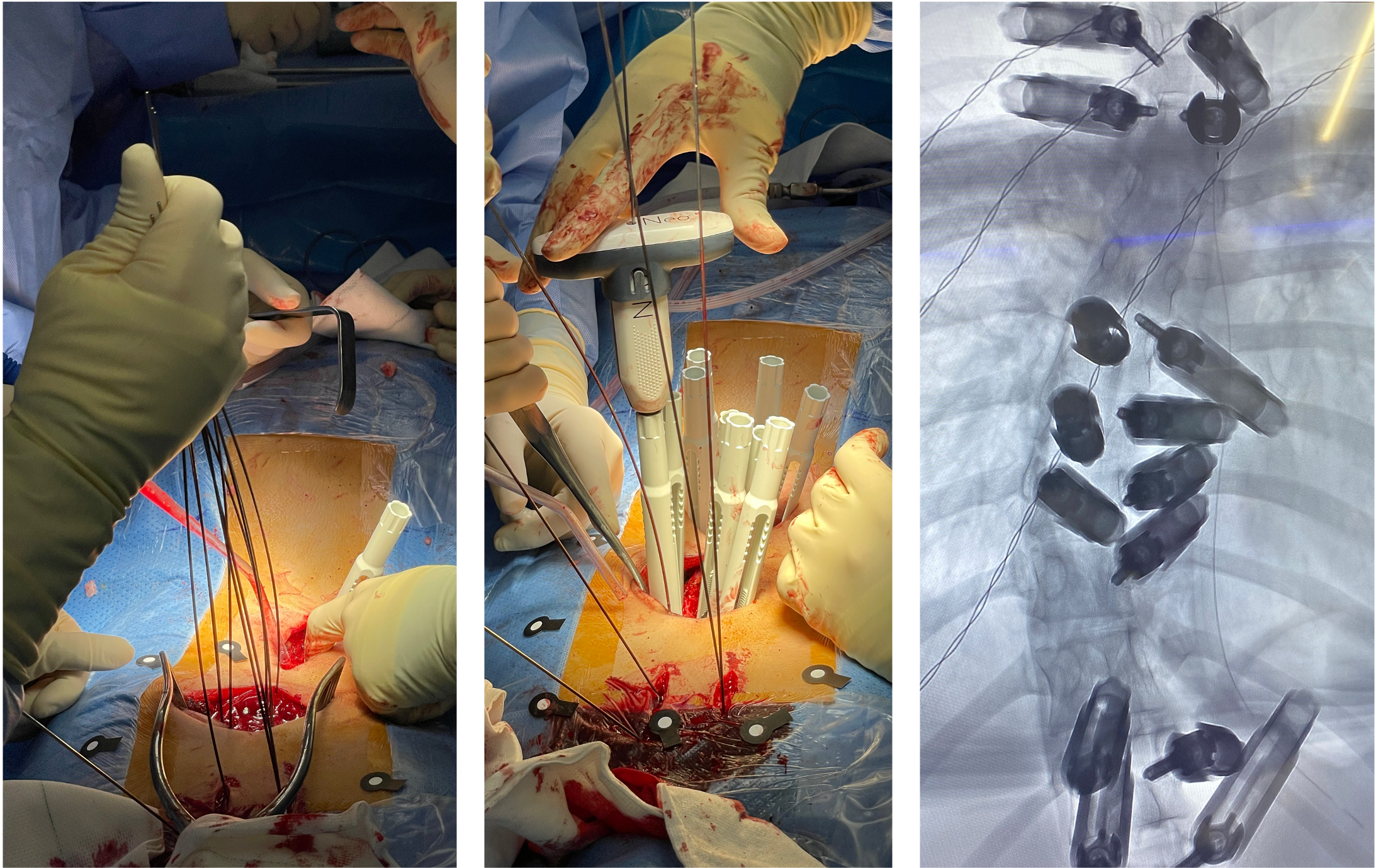
Skin incision according to plan
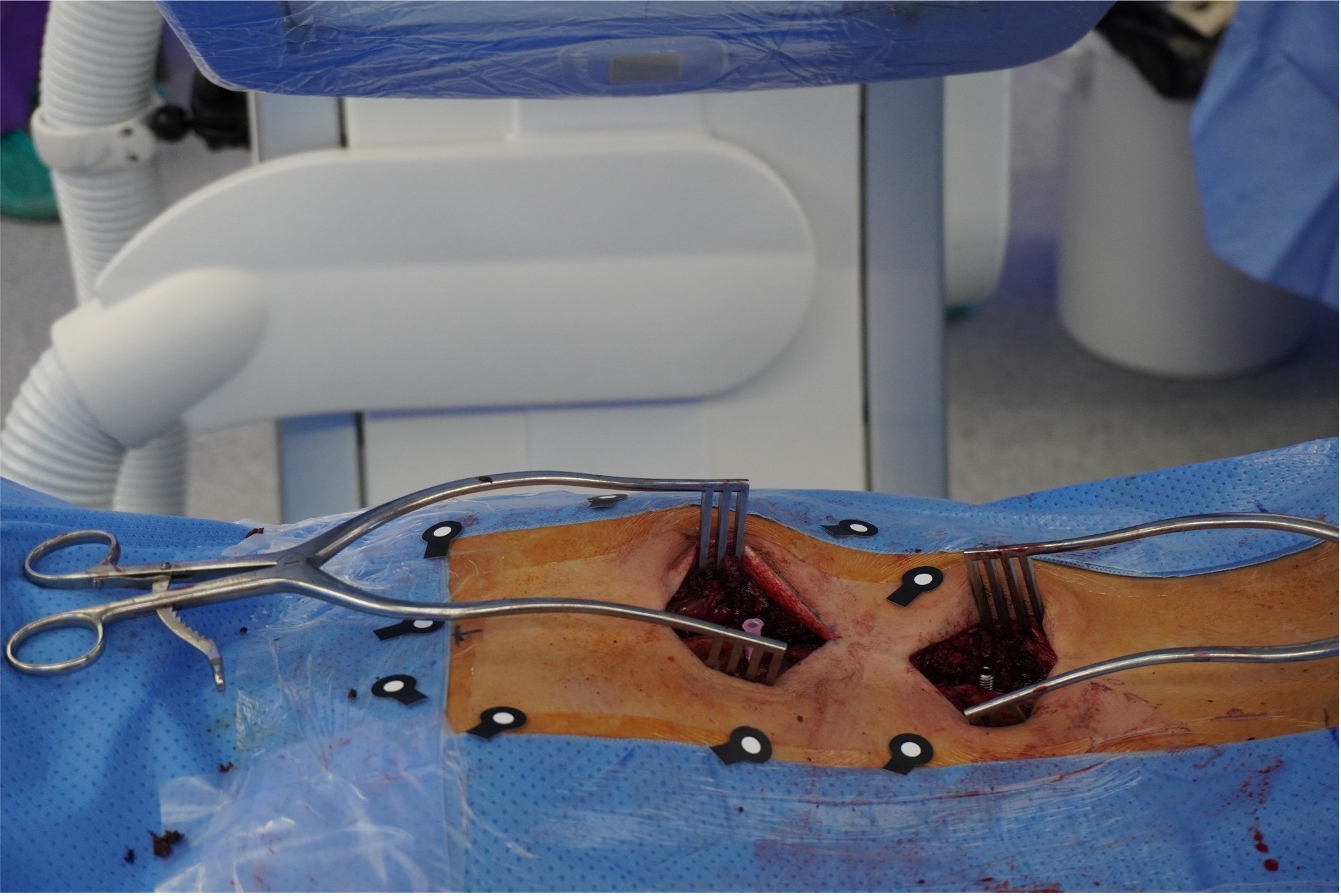


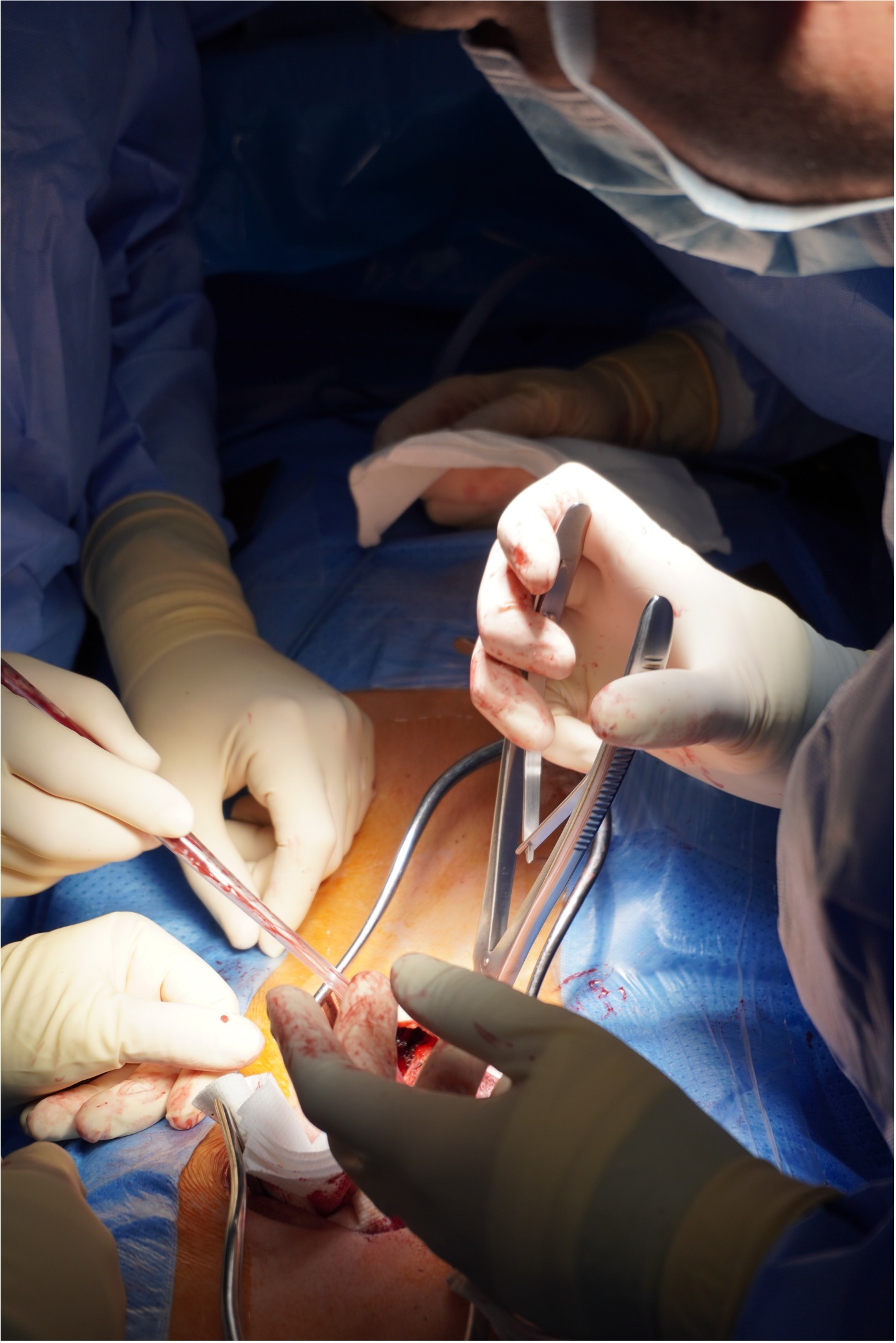
- Top – None
- Apex – Schwab Grade 1 or 2*
- Bottom – Schwab Grade 1 or 2*
* Schwab 1 = Partial facetectomy
Schwab 2 = Complete facetectomy


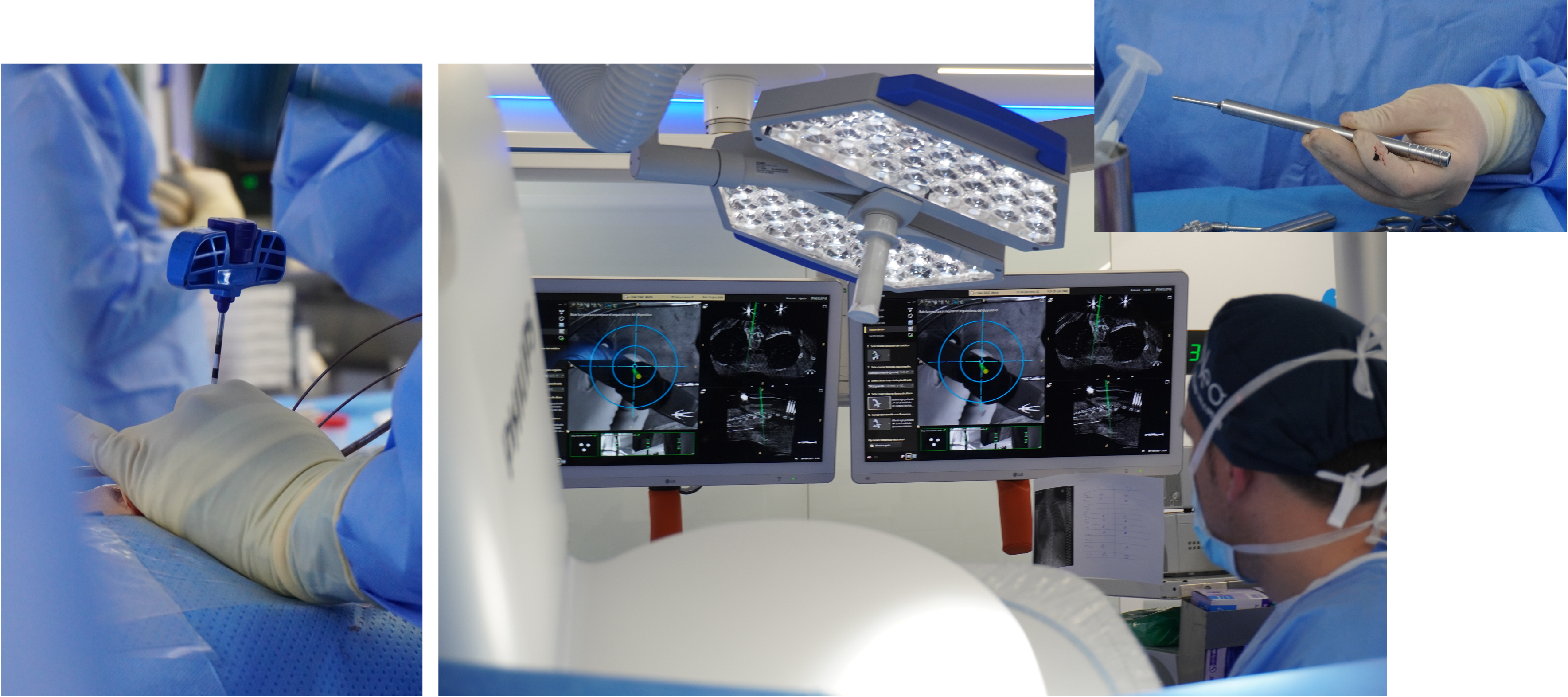


- High thoracic (T3-T4) MIS => Polyaxial
- Apex (T7 to T10) Midline open => Monoplanar
- Bottom (L1-T12) Wiltse => Polyaxial
Correction will be applied by the integrated reduction threads and spreading the correction forces over the whole construct. This is reinforced to the usage of monoplanar screws in the apex of the curve.


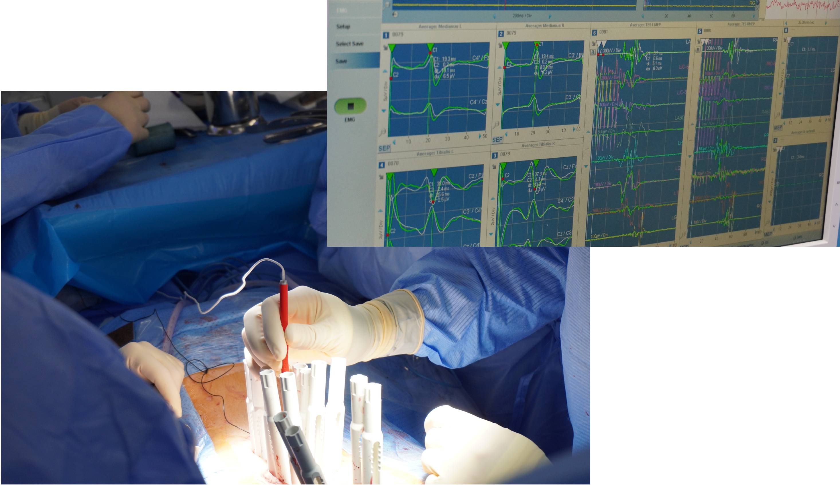
Neuromonitoring check


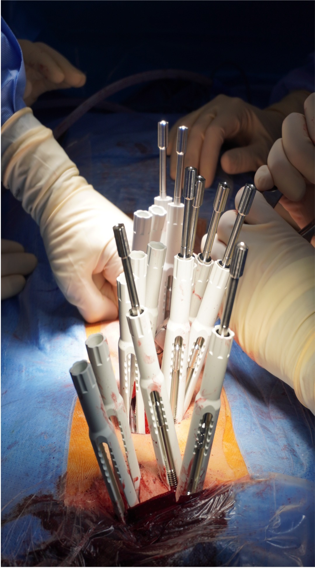
- Insertion of the CoCr rod in the concave side, set in the sagittal plane.
- Tightening of the polyaxial screws using the torque limiter at top & bottom, to create a bridge with 2 anchor points.
- Translation manœuvres from caudal to cranial with torque limiter to smoothly lift the spine up to the rod and derotate the apex using the capacity of the the monoplanar screws.
- Reinforce tightening without torque limiter
- Insertion of the bent CoCr rod in the convex side
The rod holder is a key instruments to allow for proper rodinsertion.

Post OP
Post OP results
After both sides have been tightened, the coronal alignment can be further restored by the use of the monoplanar screw guides to derotate the spine.
| Pre op | Post Op | |
|---|---|---|
| Cobb angle | 50° | 9° |
| Thoracic Kyphosis (TK) | -5° | 12.5° |
| Lumbar Lordosis (LL) | 47.3° | 45.5° |

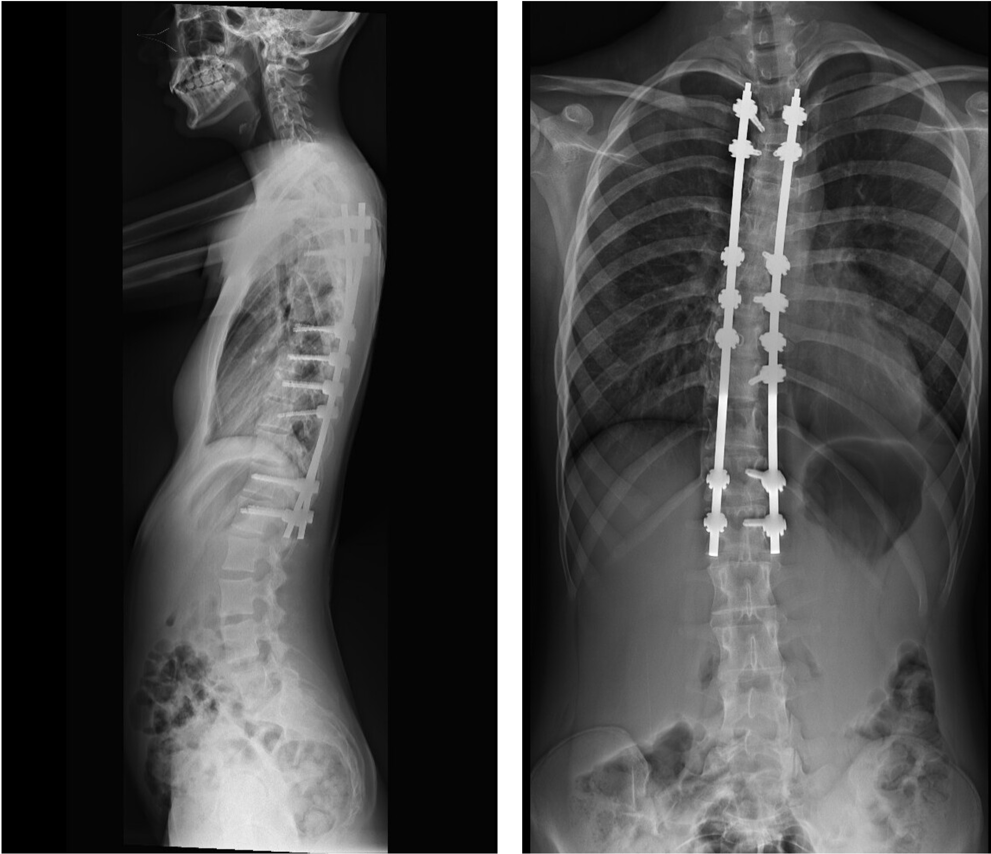
Post OP Radiographs, standing sagittal & frontal

Published with the approval of
Dr. Alejandro Peiró García


