E-Case report
Osteoporotic Fractures
Posterior long segment fixation T8 – L3Dr. med. Patrick Weidle
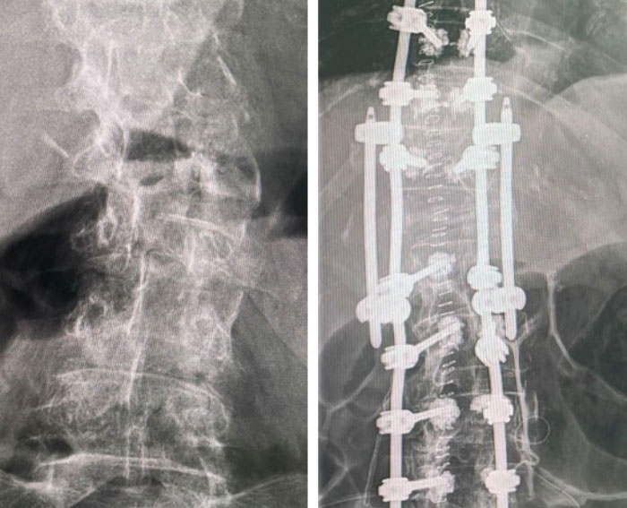
Pre OP

Clinical Case – Osteoporotic Fractures
Posterior long segment fixation T8 – L3
Dr. Patrick A. Weidle
Head of Musculoskeletal – Center MG
Head of Department for Orthopaedics-, Trauma-, Spine Surgery and Interventional Pain Therapy
Krankenhaus Neuwerk
Mönchengladbach
Germany


Patient Information
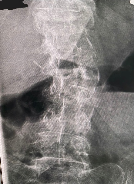
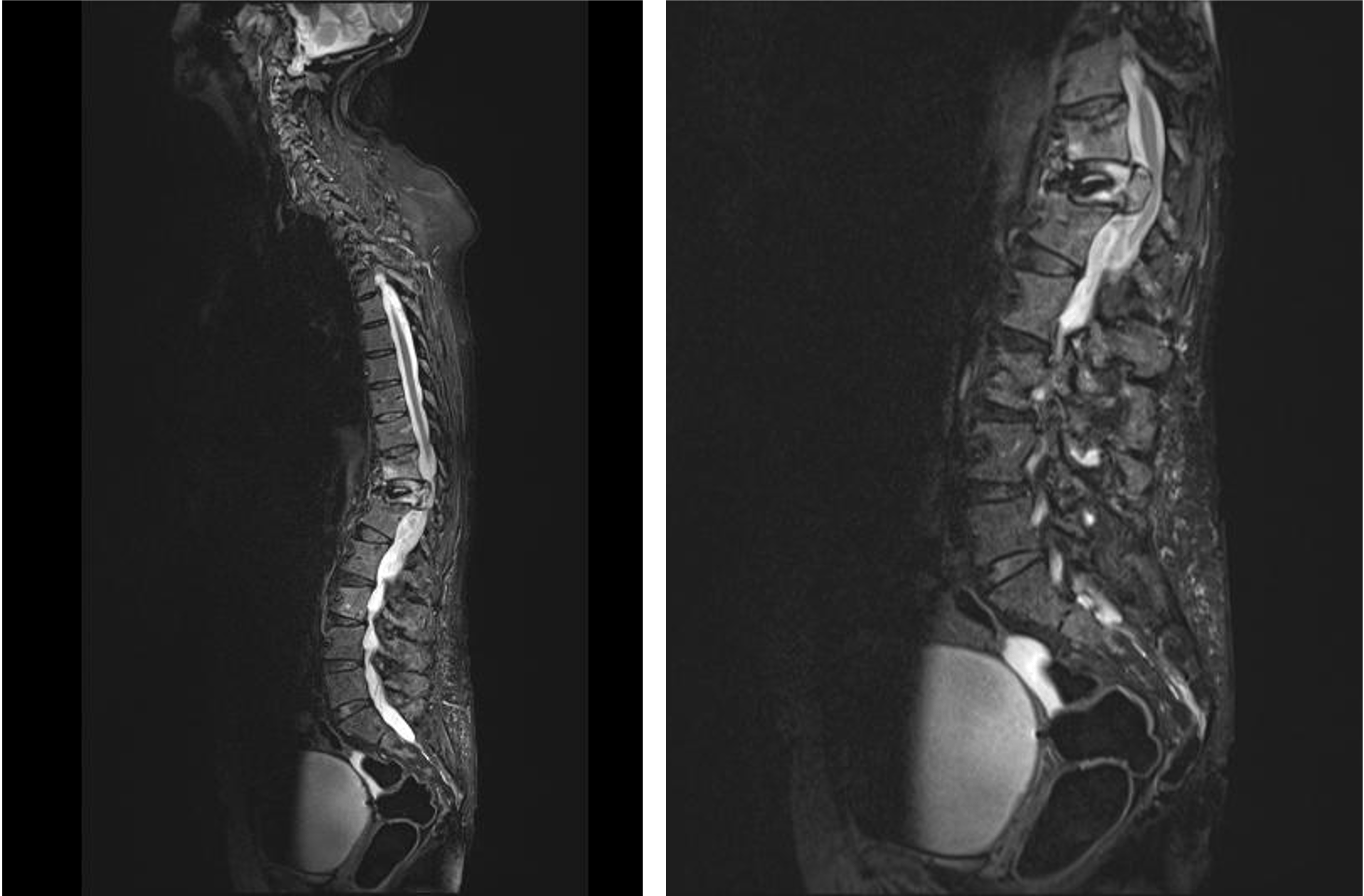
Female 80 years old patient suffers from osteoporotic fractures in the thoraco-lumbar junction area, T10, T11 and L2.
Diagnoses:
- T10 – OF1-FX
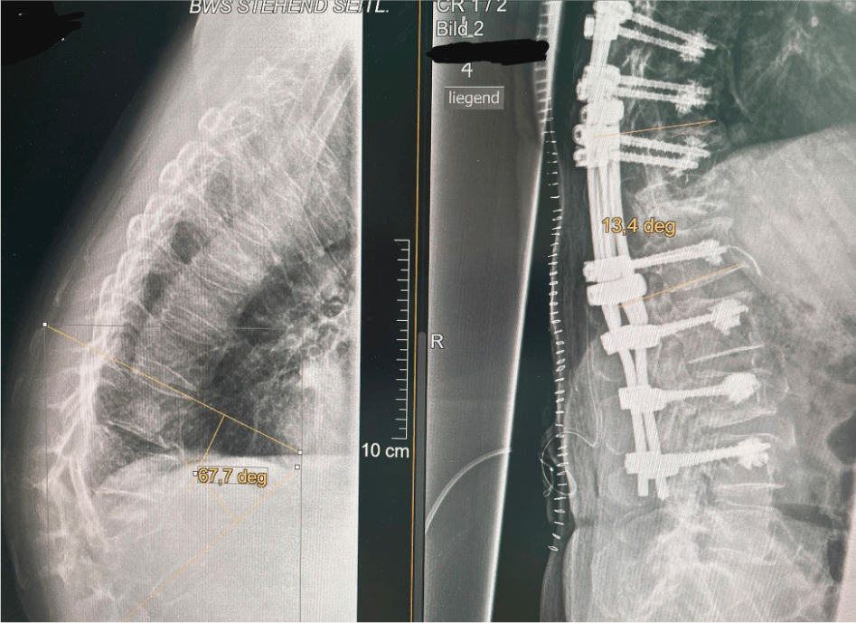
- T11 – OF5-FX – Kyphosis T10-T12 Cobb-Angle 57° with temp. Plegia
- L2 – OF1-FX
- Adult Scoliosis
- Osteoporosis – Bone density T-score: -3.5
The patient has been conservatively treated by the General Practitioner.
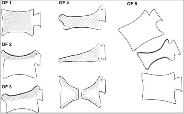
Schnake KJ, Blattert TR, Hahn P, et al. Spine Section of the German Society for Orthopaedics and Trauma. Classification of Osteoporotic Thoracolumbar Spine Fractures: Recommendations of the Spine Section of the German Society for Orthopaedics and Trauma (DGOU). Global Spine J. 2018 Sep;8(2 Suppl):46S-49S.


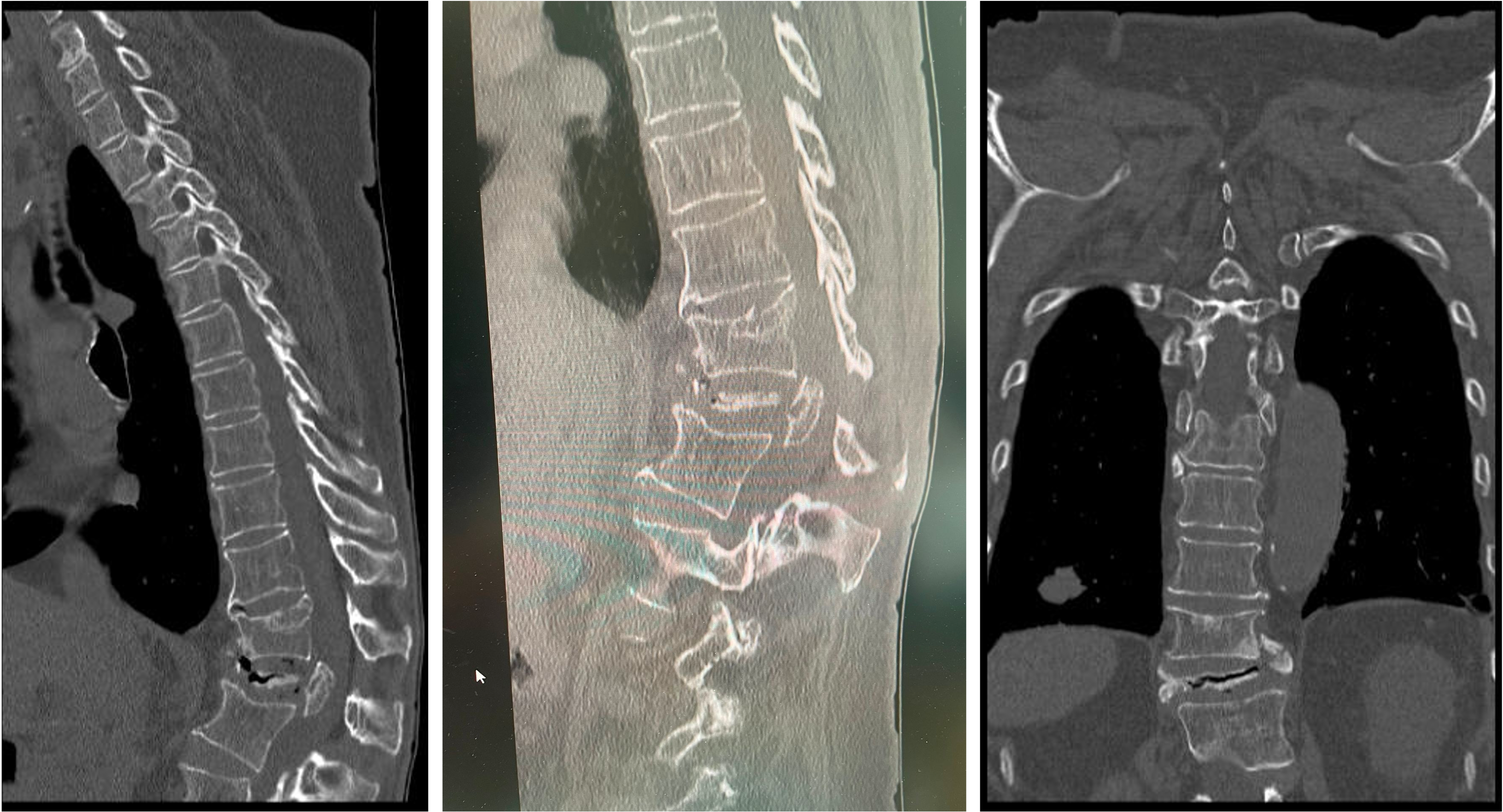
Pre OP CT and radiograph,
sagittal & coronal views

Pre OP radiograph coronal & MRIs sagittal


OR-Strategy:
- Open spondylodesis T8-L3 in IONM and 3D-navigation
- Cement augmentation of all screws
- Modified PSO T11 & reduction with AR technology
- Quattro rod enhanced fixation with parallel 4 connectors
Secondary surgery:
- TT-VBR T11

Intra OP

- Posterior long segment fixation with the Neo Pedicle Screw System™
- Totally 14 screws in T8 – L3 levels
- All pedicle screws were placed with guidance/support of navigation
- Surgery performed with help of IONM
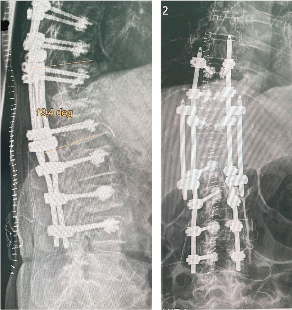
- Quattro rod enhanced the fixation with 4 parallel connectors. The satellite rods were used to achieve increased stability.
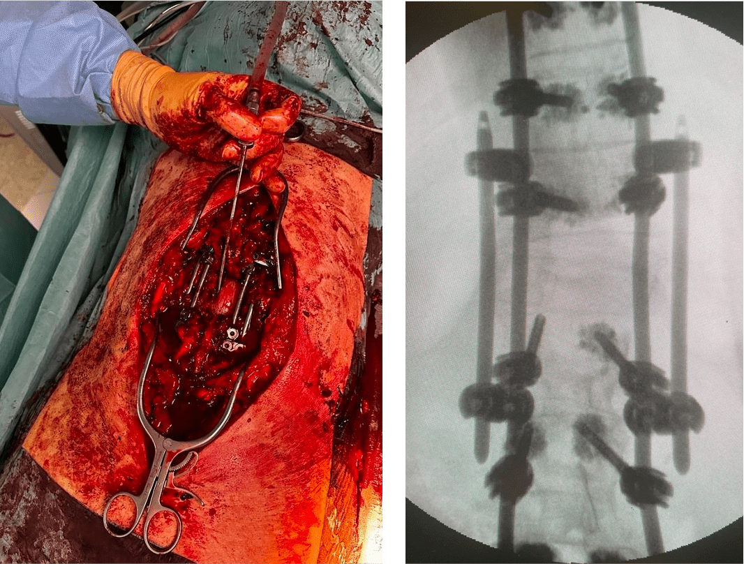

- All screws were cement augmented because of the poor bone quality.
- Correction supported by Schwab 2/3 modified PSO
- AR supported technology was used to objectify the amount of the intraoperative correction: From 67° to 13.4° at the thoracolumbar junction.
- Vertebral Body Replacement T11 will follow in 2nd surgery.

Post OP

Post OP radiographs,
sagittal & coronal views

Published with the approval of Dr. Patrick Weidle


