E-Case report
Degenerative Scoliosis
Fusion & Posterior fixation L2 – S1Thomas Schachtschabel, MD

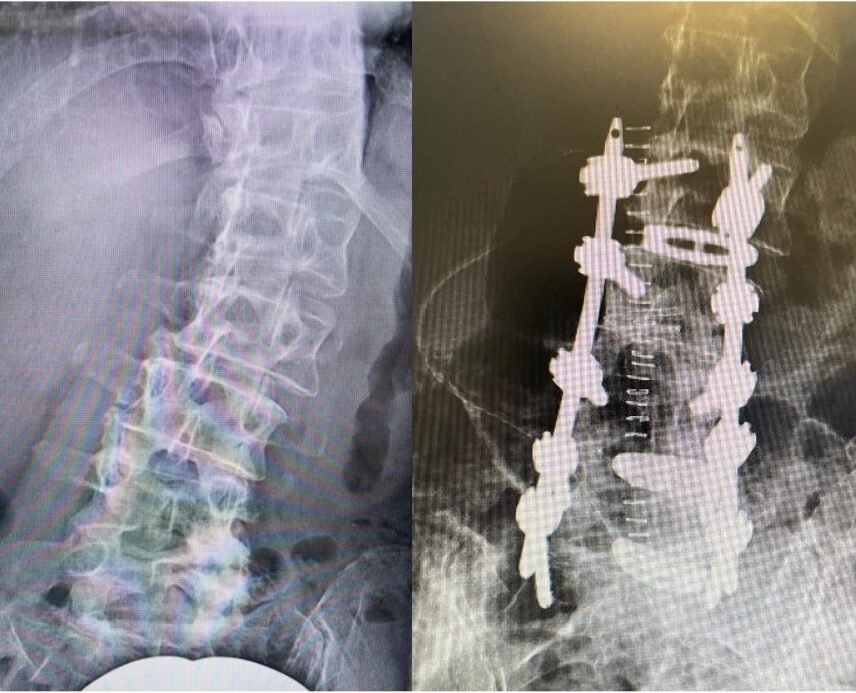
Pre OP

Clinical Case – Degenerative Scoliosis
Fusion & Posterior fixation L2 – S1
Neo ADVISE™
Dr. med. Thomas Schachtschabel
Neurosurgeon
ORTHO CENTRUM SAALE
Bad Neustadt Saale/Gersfeld
Germany

Patient information
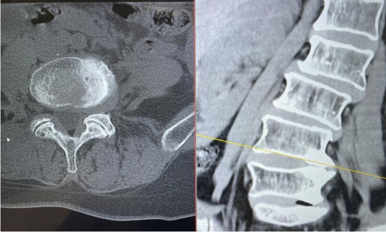
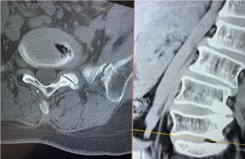
Female 60-year-old patient diagnosed with degenerative deformity of the lumbar spine.
Slipped disc in L2/3 right side.
In levels L4 – S1 left side slipped disc, osteochondrosis and foraminal stenosis.
The patient suffers from pain on the right side to the hip and thigh, and in her left side to the bunion.
Bone quality is normal for her age.
Planned Surgery:
-
Percutaneous MIS surgery
-
Intervertebral fusion TLIF L2/3, L4/5, L5/S1
-
Autogenous bone graft
-
Posterior fixation L2 –S1
-
Neo ADVISE™ to be used


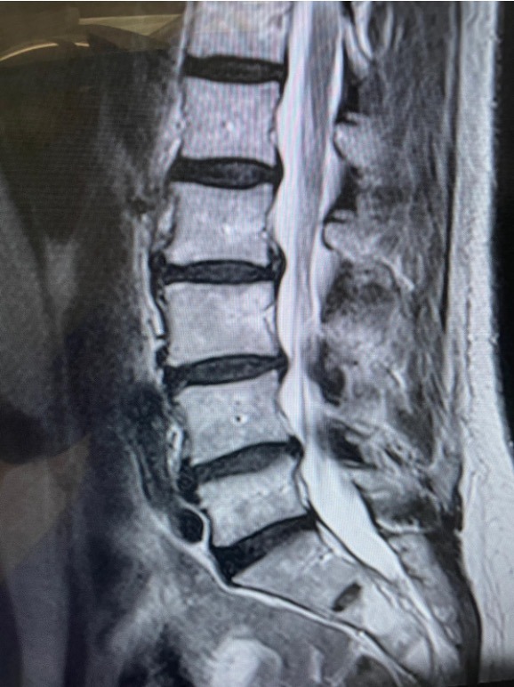
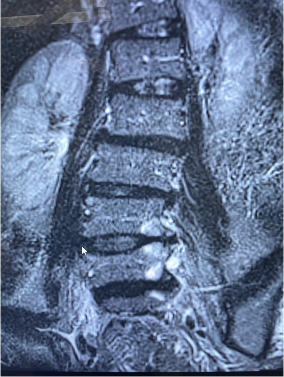
Pre OP radiographs & MRI Sagittal & frontal views
Pre OP radiographs & MRI
Sagittal & frontal views
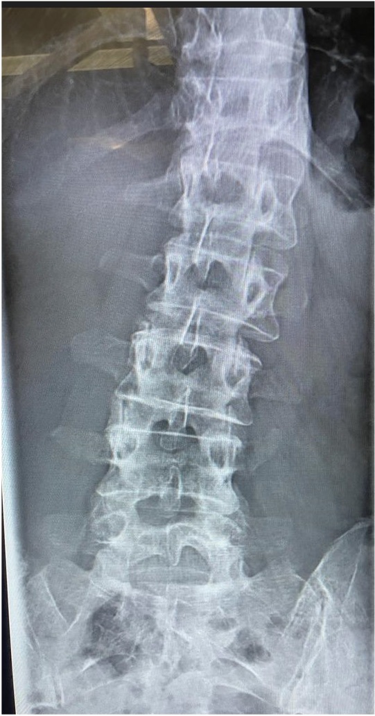
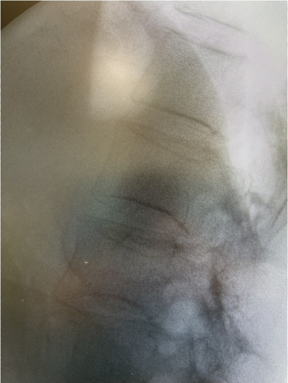


Pre OP CTs

Intra OP

L2/3 – Placing K-wires, midline/ transmuscular approach, preparation of intervertebral space, cage insertion right, Cage anatomical/curved 32mm, 9mm, 5°
Intervertebral fusion using the Neo Cage System™
Totally 3 Neo TLIF anatomical curved cages were inserted.
Autogenous bone graft was used.
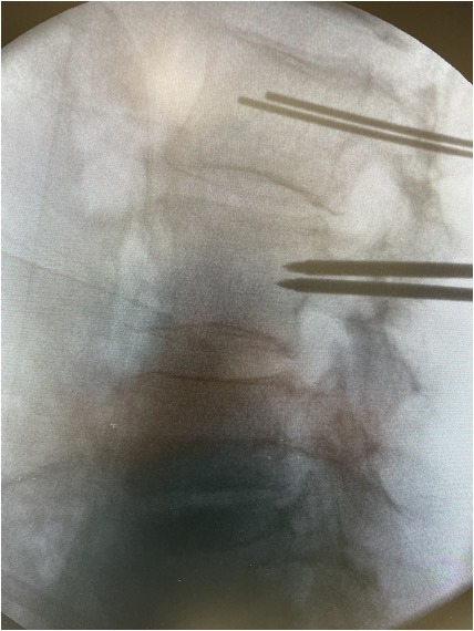
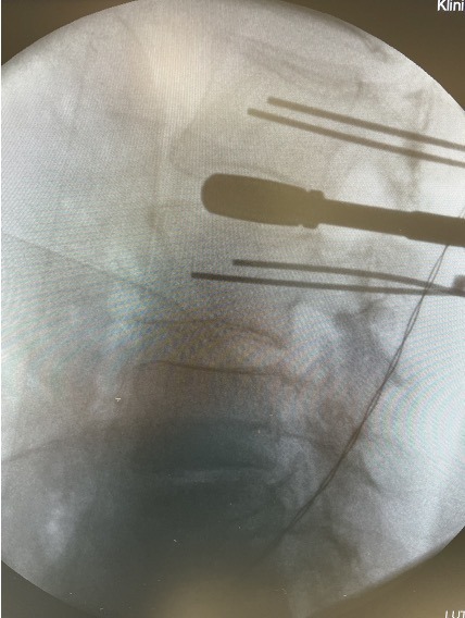

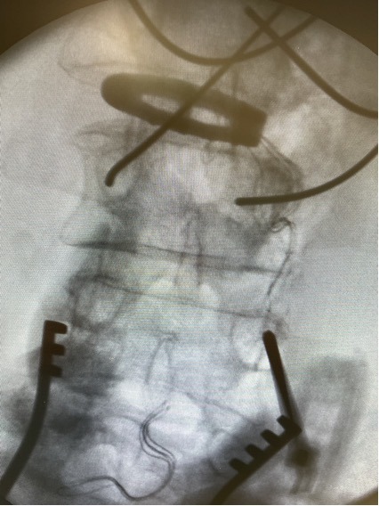
L2/3 – Placing K-wires, midline/ transmuscular approach, preparation of intervertebral space, cage insertion right, Cage anatomical/curved 32mm, 9mm, 5°


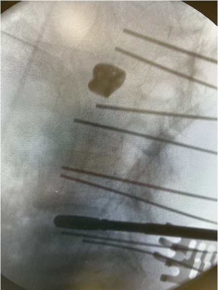
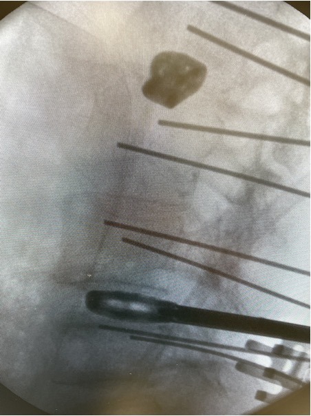

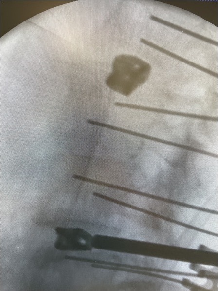
L4/5 -preparation of intervertebral space, cage insertion left. Cage anatomical/curved 32mm, 9mm, 5°


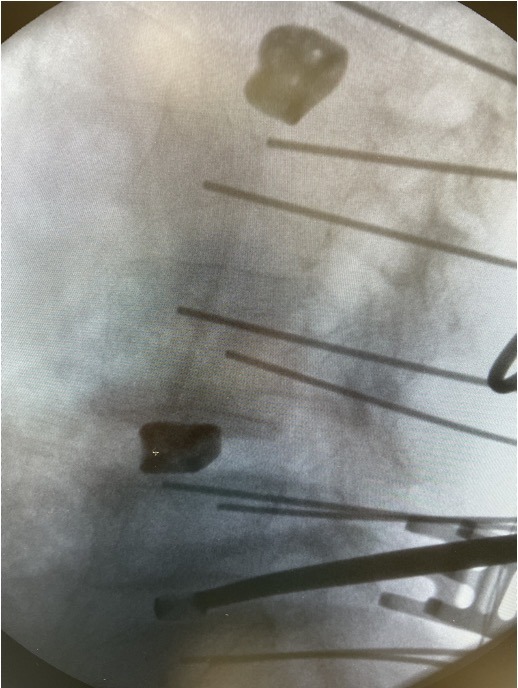

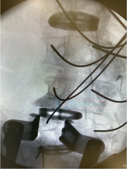
L5/S1 -preparation of intervertebral space, cage insertion left. Cage anatomical/curved 32mm, 7mm, 5°


Posterior stabilization with the Neo Pedicle Screw System™
Totally 10 pedicle screws placed over the levels L2 – S1.
- L2 – 2 x Ø5.0×45 mm
- L3 – 2 x Ø6.0×45 mm
- L4 – 2 x Ø6.0×45 mm
- L5 – 2 x Ø6.0×45 mm
- S1 – 2 x Ø6.0×40 mm


Usage of the Neo ADVISE™
Usage of the Neo ADVISE™
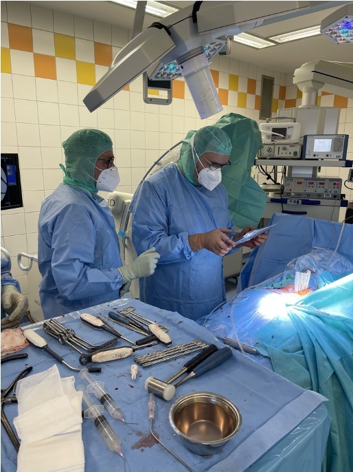
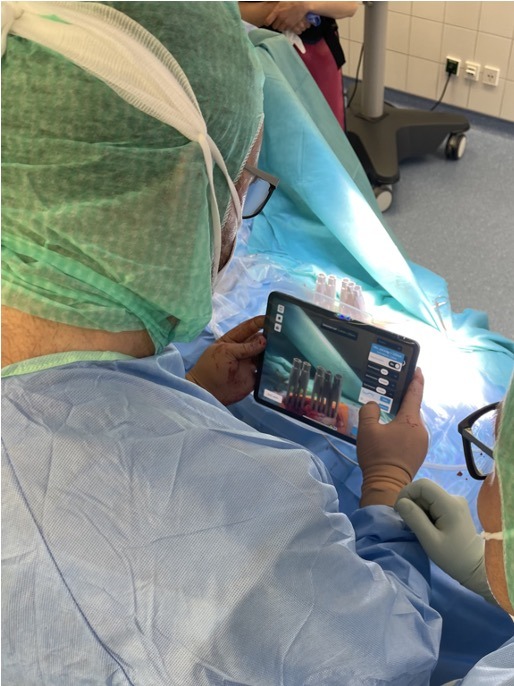
Scene analysis and mapping of the guide towers by using the iPad screen

Confirming the rod shape.
The lenght of this rod is 130mm.
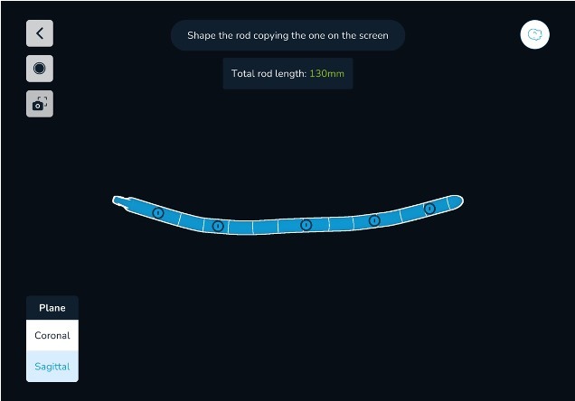
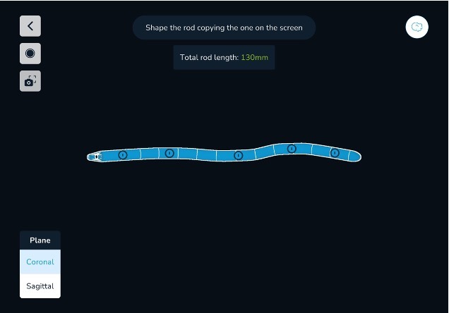
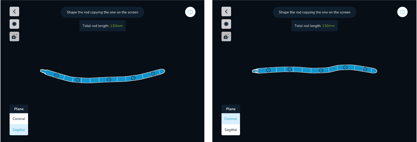
Visualizations of the shape of the rod, in both the sagittal and the coronal planes shown in the iPad screen.

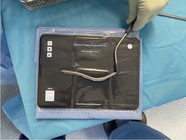
The second rod has been bent in the sagittal plane
Bending of the rod, 130mm straight, by using the visualization of the rod in thesagittal and coronal planes shown in the iPad screen.

Rod placement
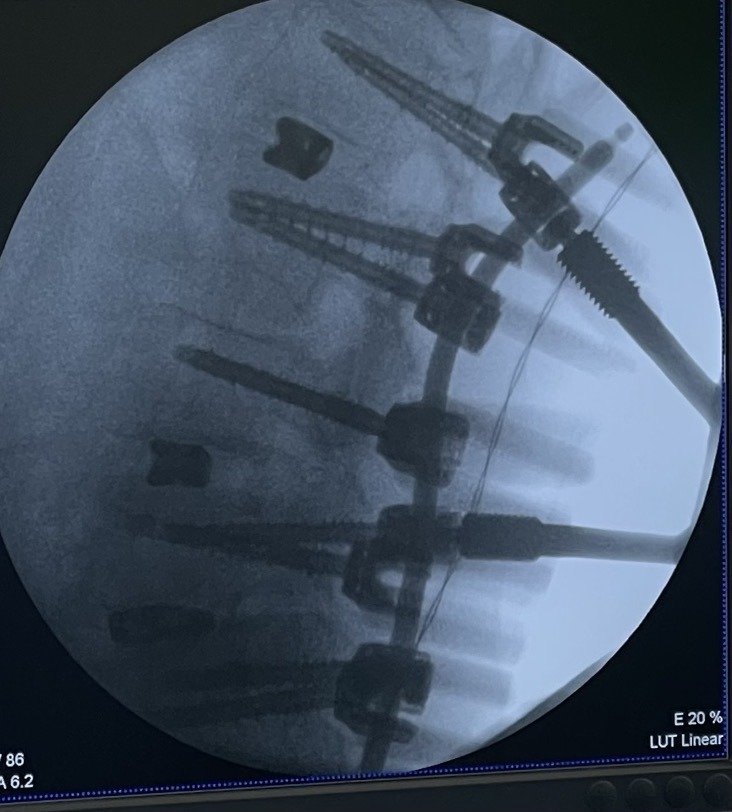


Pre-fixation of the set screws, using the torque limiter attached to the handle, final tightening of all set screws and removal of all guide towers.

Post OP

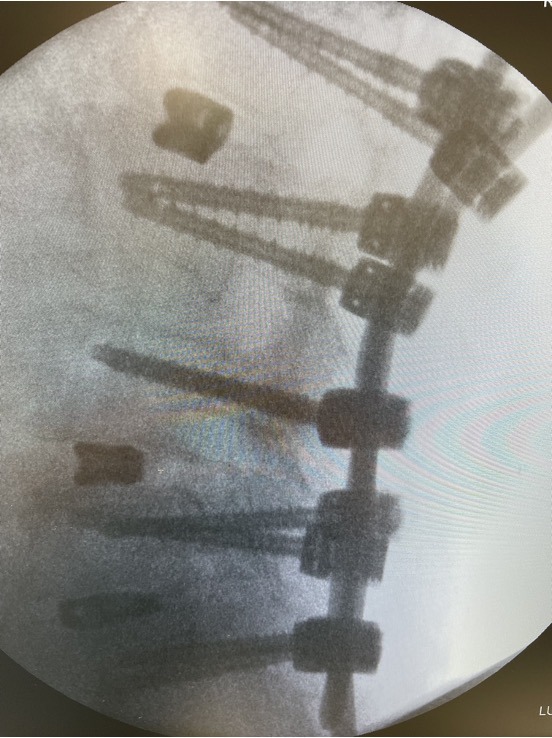
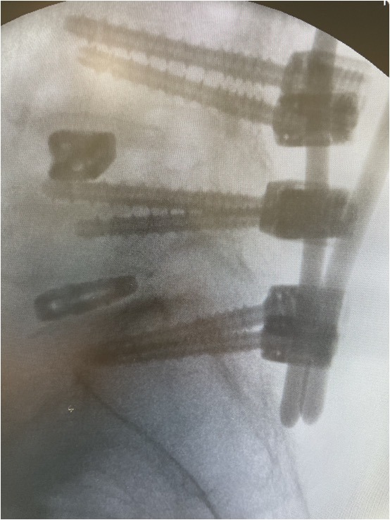
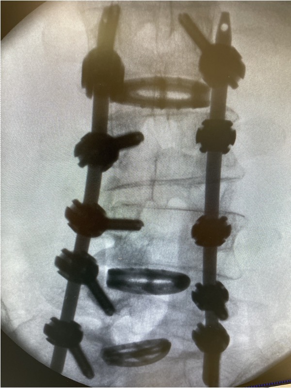
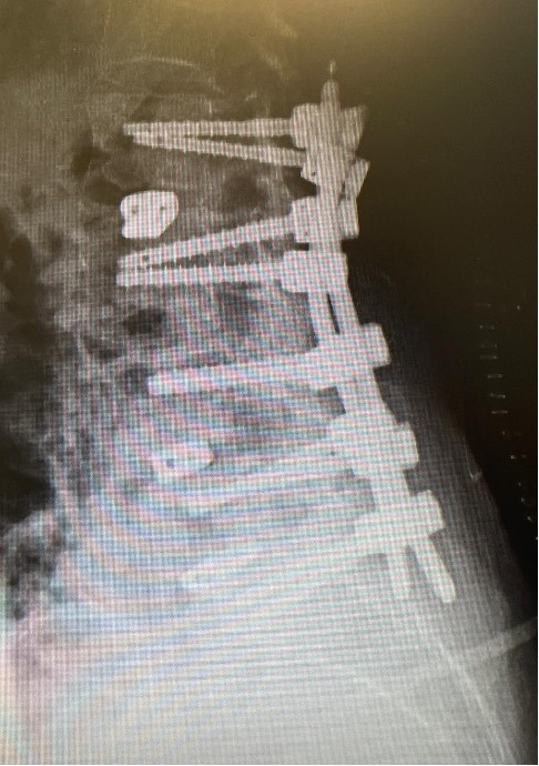
Radiographs, after final fixation of the posterior construction and removal of the instrumentation.
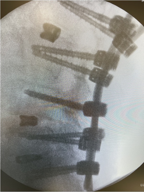
Radiographs, after final fixation of the posterior construction and removal of the instrumentation.


Post OP radiographs, standing
- Intra OP Blood loss: 200 ml
- Duration of surgery: 210 min
- Patient was discharged from the hospital after 7 days
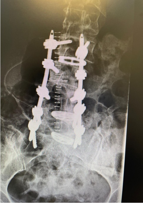

Published with the approval of Dr. Thomas Schachtschabel


