E-Case report
Disc herniation & bilateral foraminal stenosis
L4/5 MIS TLIF in ASC outpatient procedureAli M. Maziad, MD PhD

Pre OP

Clinical Case – Disc herniation & Bilateral foraminal stenosis
L4/5 MIS TLIF in ASC* outpatient procedure
Ali M. Maziad, MD PhD
Orthopedic Spine Surgeon
Maziad Spine Institute
Miami, FL, USA


*Ambulatory Surgical Center


Patient Information:
35-year-old male patient bodybuilder with chronic low back pain (LBP) and a recent history of motor vehicle accident. Left lower extremity radicular pain.
- Height: 71.0 in/1.80 m
- Weight: 250 lbs/113 kg
- BMI: 34.9 / BSA: 2.3
Diagnoses:
- L4/5 disc herniation
- Bilateral foraminal stenosis with retrolisthesis and instability on flexion-extension view
- No radiculopathy


Pre OP radiographs, frontal and sagittal views


Pre OP MRI and radiograph sagittal view

Pre OP MRIs sagittal and axial views


Intra OP
OR-strategy:
- Thoraco-Lumbar Interfascial Plane (TLIP) block, injection of local anesthetics, in L3 – L4.
- L4/5 minimally invasive TLIF – left, to be performed as an outpatient procedure in the ASC.
- Planning to use the Neo Cage System ™ & Neo Pedicle Screw System™ for fusing the segment L4/5.
Intra OP radiograhs for the planning of the surgical access.


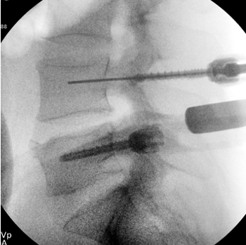
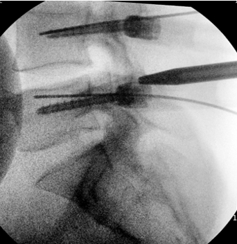
L5 - Access and placing the K-wire, left side


L4 - Access and placing the
K-wire, left side


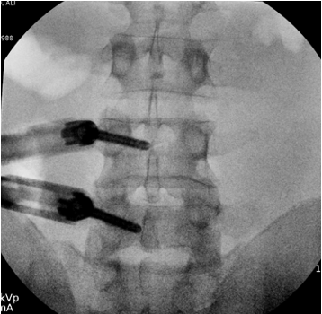
Inserting at the left side 2 pedicle screws,
Neo Pedicle Screw System™
L4: Ø6.0x45 mm
L5: Ø6.0x45 mm
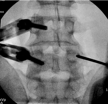
Access and placing K-wires, right side
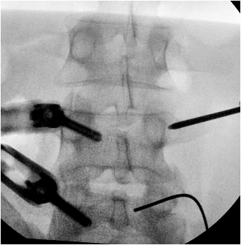
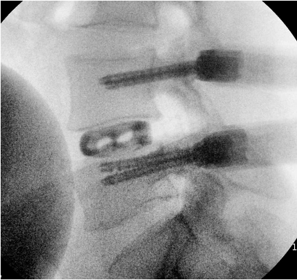
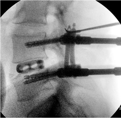
Disc space preparation and placement of Neo Cage anatomical/straight 32mm, 12mm, 0°


Placement of the Neo Cage


After the placement of the Neo cage, another 2 Neo pedicle screwswere inserted at the left side,
L4: Ø6.0x45 mm
L5: Ø6.0x45 mm
2 X titanium rods, straight 40mm/45mm, were placed.
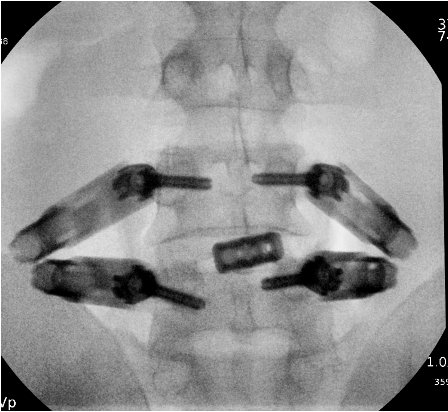
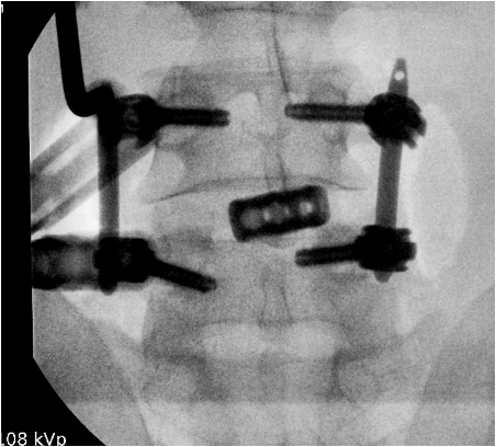

Radiographs, frontal and sagittal views
Total time of surgery: 2 hours and 10 minutes
Estimated blood loss: 15 cc


Post OP
Radiographs after the final fixation of the posterior construction, and removal of the instrumentation
Post OP radiographs, 1 week after the surgery, frontal and sagittal views.
Patient was discharged from the clinic 1 hour after surgery.
He consumed minimal narcotics on the first 48 hours.
Pain resolved afterwards and he used Tylenol as needed.
Reported resolution of lower back pain and no radicular pain 1 week postoperatively.

Follow up
Radiographs at 6 weeks Follow Up, frontal and sagittal views


Radiographs after the final fixation of the posterior construction, and removal of the instrumentation
Follow up
Radiographs at 3 months Follow Up, frontal and sagittal views


Post OP radiograph at 5 months FU confirms the stable clinical result.

Picture showing the nicely healed minimal incisions at the lower back of the patient.

Published with the approval of Dr. Ali Maziad