E-Case report
Bechterew’s disease ankylosing spondylitis (AS) in T7/8
Posterior fixation T5 – T10 Stefan Piltz, PhD

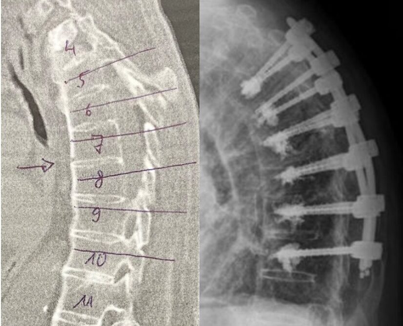
Pre OP

Clinical Case – Bechterew’s disease ankylosing spondylitis (AS) in T7/8
Posterior fixation T5 – T10
Neo ADVISE™ fixation module
Prof. Dr. med. Stefan Piltz
Head of Trauma Department
Orthopaedic/ Trauma Surgeon
Klinikum Coburg
Coburg, Germany


Patient Information
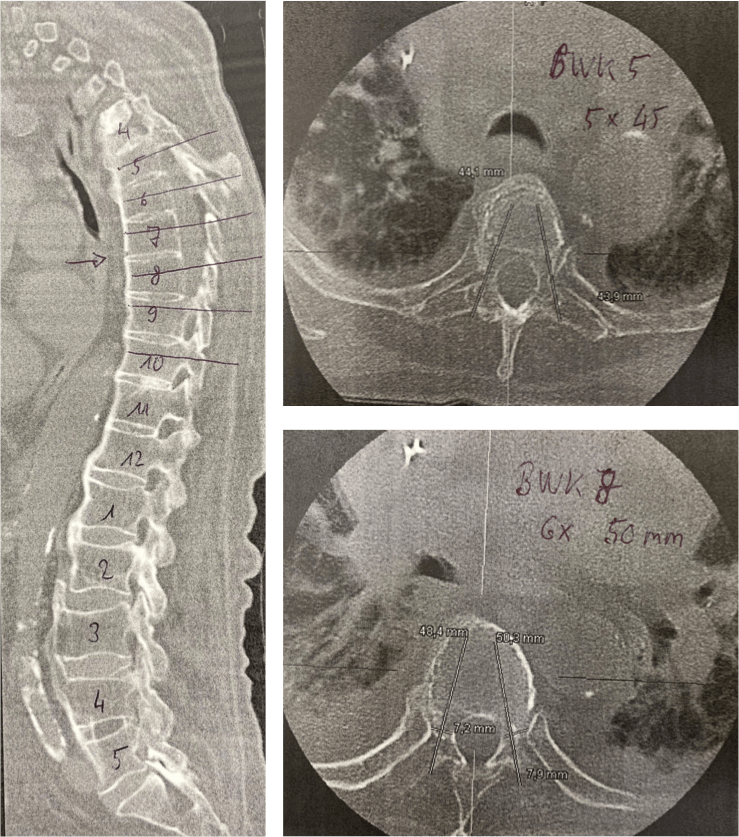
Male 90 years old patient diagnosed with ankylosing spondylitis (AS) in level T7/8.
- Bechterew’s disease
- Poor bone quality
- Reduced lung function
- Poor kidney values
Planned Surgery:
- General Anestesia
- Posterior fixation T5 – T10
- Minimally Invasive Surgery
- Percutaneous access
- Neo ADVISE™ application to be used


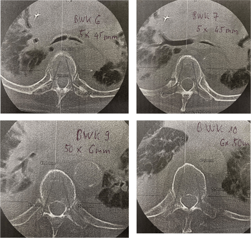
Pre OP CT sagittal & axial views

Intra OP

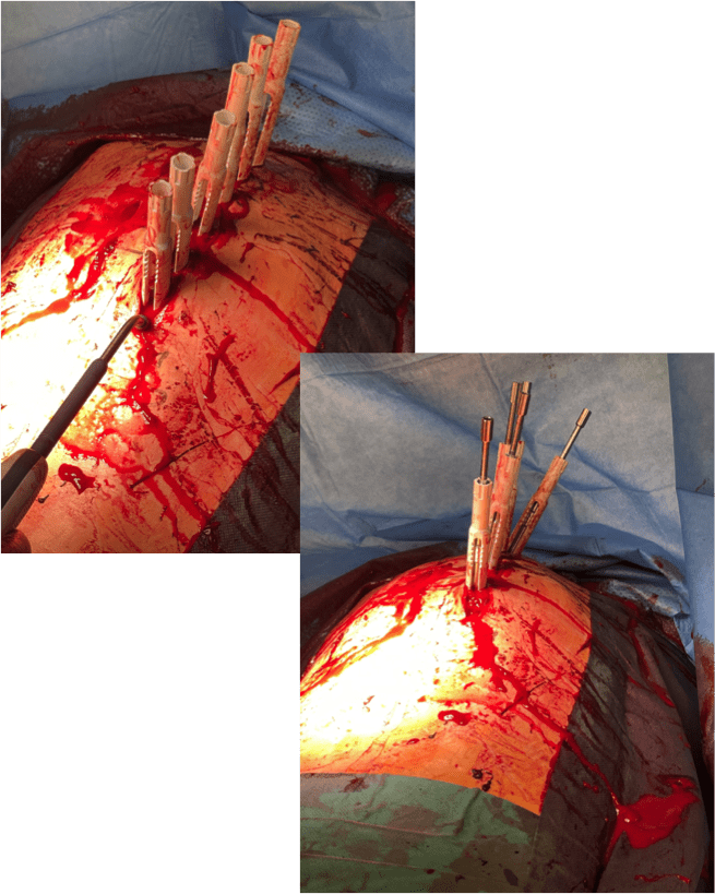
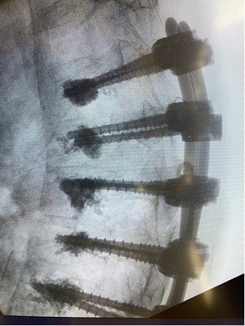
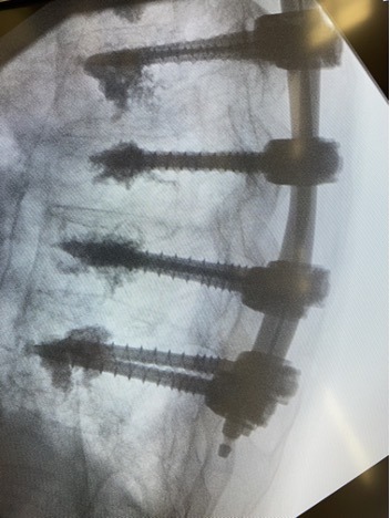
Posterior fixation with Neo Pedicle Screw System™
Totally 12 pedicle screws placed over the 6 vertebral levels T5 – T10
T5 – 2 x Ø5.0 x 45 mm
T6 – 2 x Ø5.0 x 45 mm
T7 – 2 x Ø5.0 x 45 mm
T8 – 2 x Ø6.0 x 45 mm
T9 – 2 x Ø6.0 x 50 mm
T10 – 2 x Ø6.0 x 50 mm
2 x 160 mm straight titanium rods
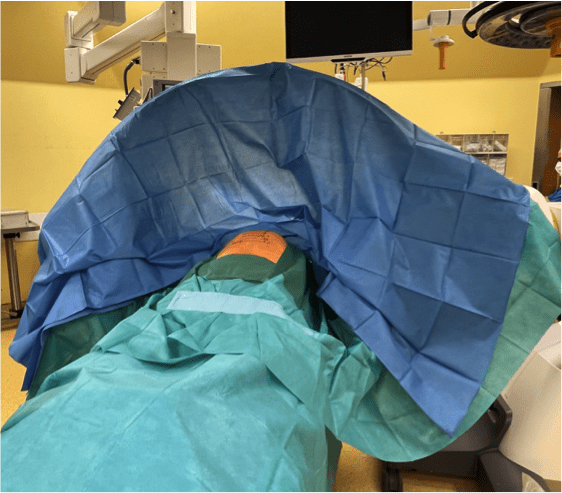
Preparing for the surgery
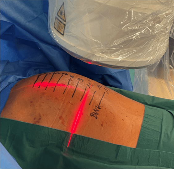


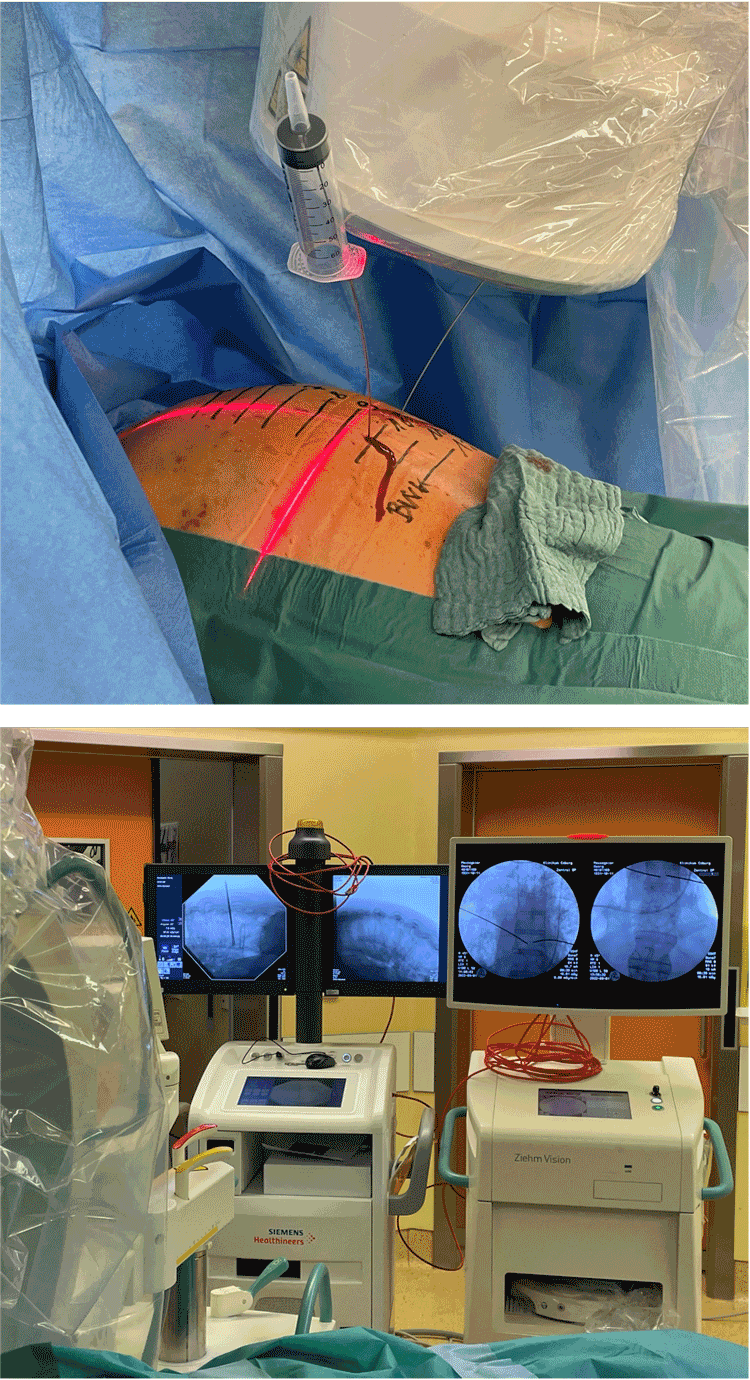
K-wires to guide the placement of the pedicle screws
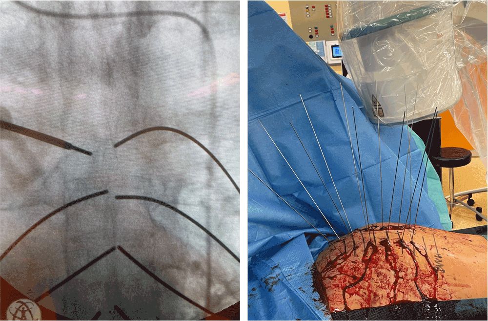


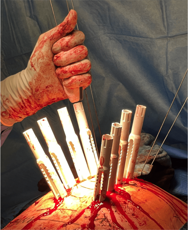
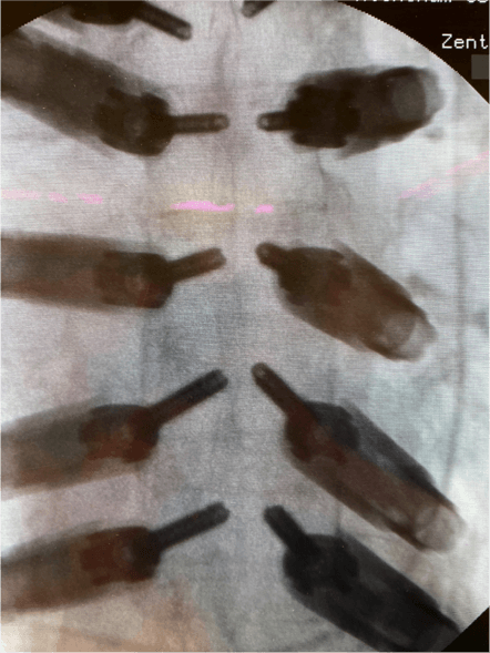
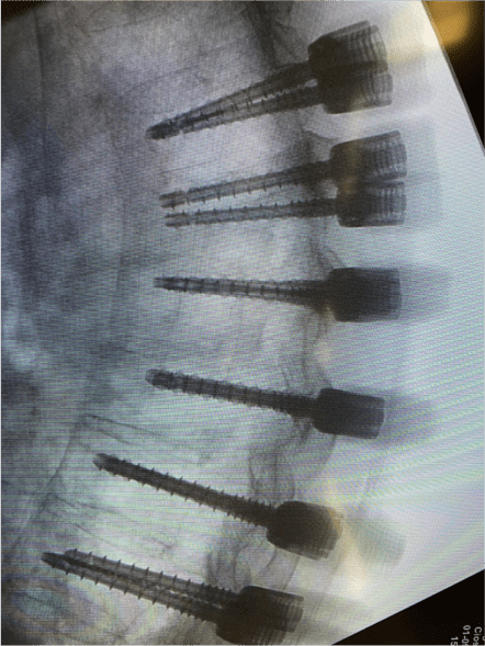
Pedicle screws are placed


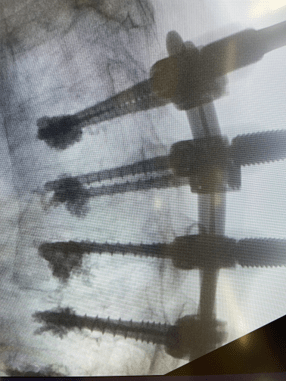
All screws are cement augmented with 1ml cement per screw injected.
Neo system pedicle screws are all cannulated and fenestrated.
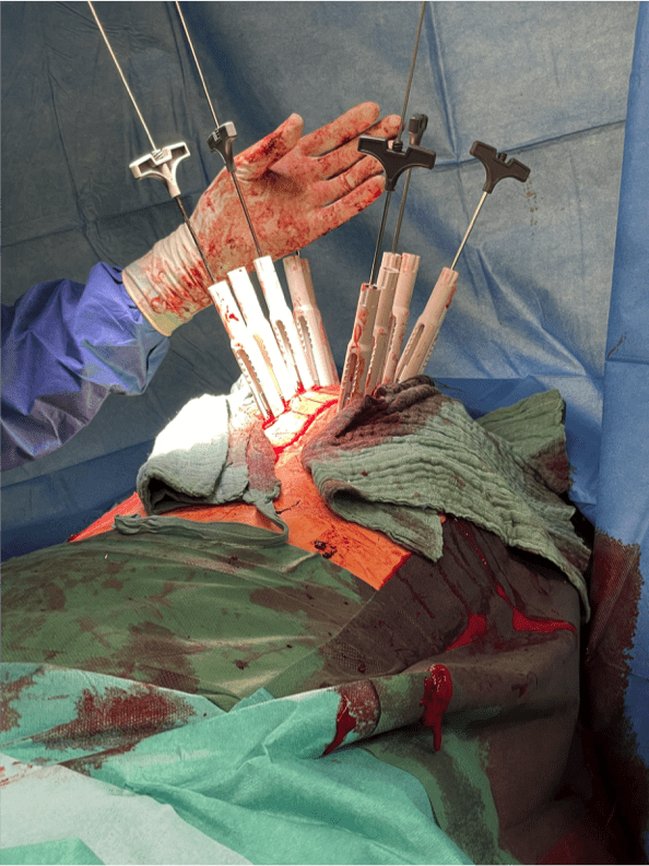
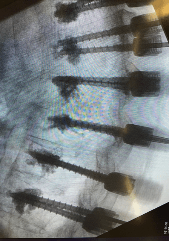

Usage of Neo ADVISE™
Scanning the screw extenders, access and analyze of the data, rod selection, and getting the visualizations of the ideal rod bending, in both the sagittal and the coronal planes, in the iPad screen.
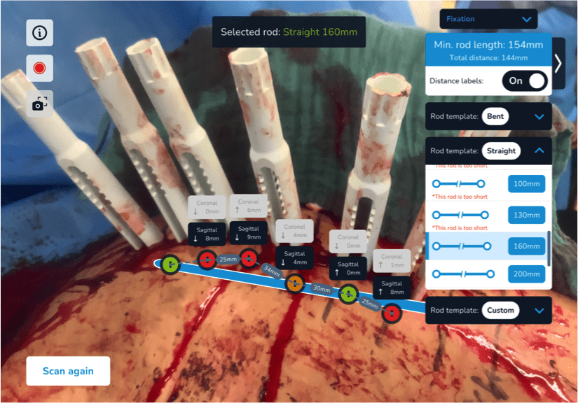
Visualizing the off-set in both planes relative to the screw heads in relation to the selected straight 160mm rod.
The screw adjustments are color-coded into the virtual screw heads. The colors green, orange, and red indicate different levels of adjustments.


Rod bending using the rod visualization in the iPad screen as template for the sagittal and coronal planes.
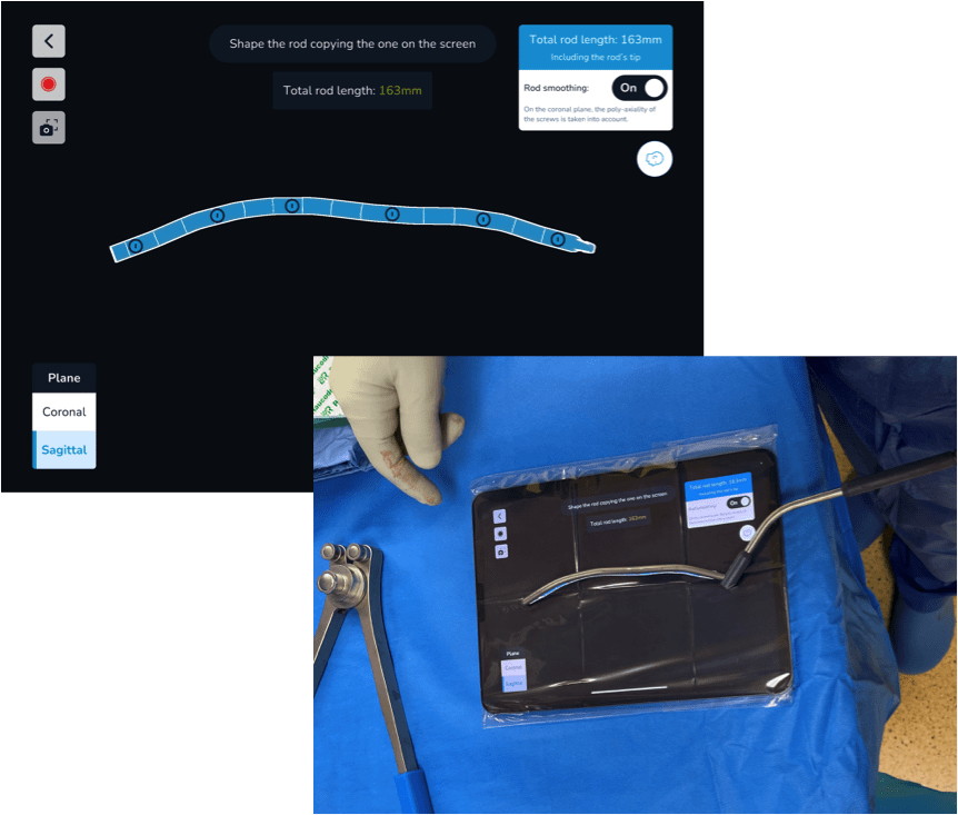
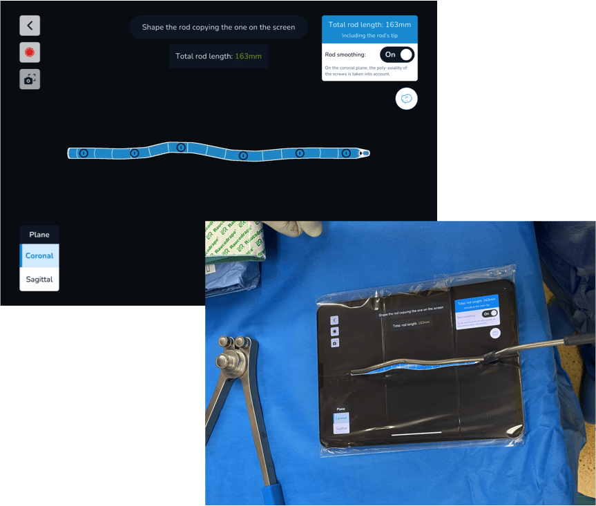


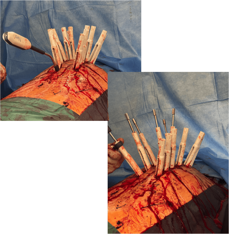
Placement of rods and final tightening of the screws, left and right sides

Post OP
Post OP
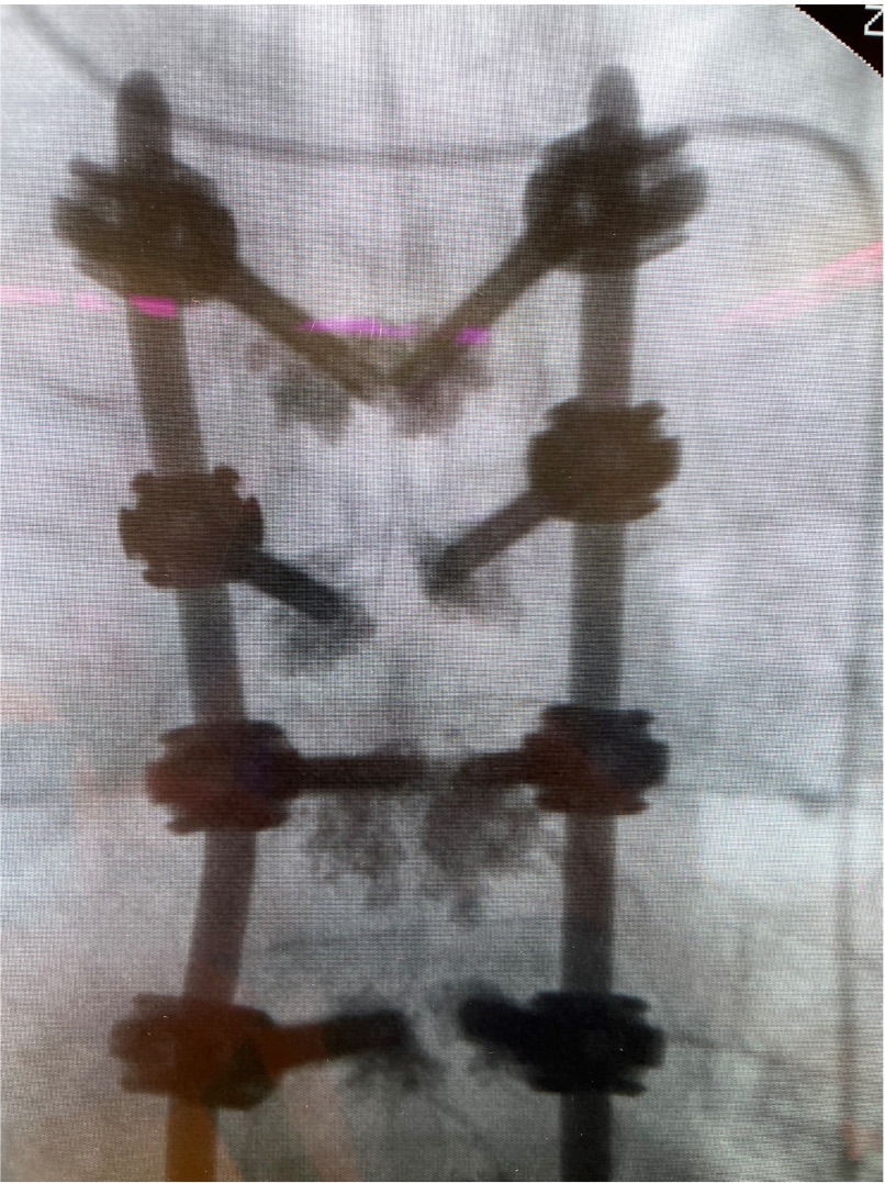
Radiographs after final fixation of the posterior construction in frontal and sagittal views
- Intra OP bloodloss: 500 ml
- Duration of surgery: 180 minutes
- Neurological status: OK
Patient was discharged from hospital after 20 days.
Follow Up visit
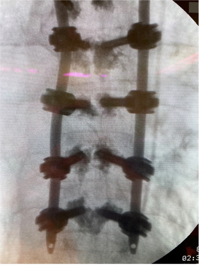
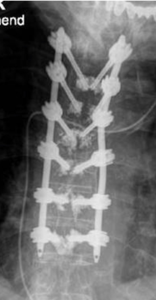
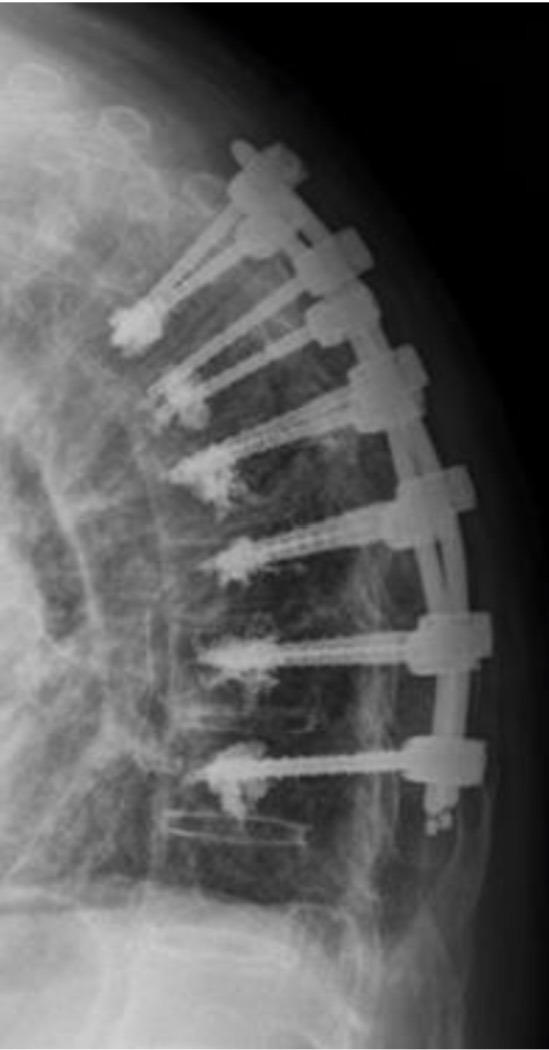
Standing radiographs were taken a few weeks after the surgery took place.

Published with the approval of
Prof. Stefan Piltz


